J Adv Prosthodont.
2017 Dec;9(6):432-438. 10.4047/jap.2017.9.6.432.
Detection of proximal caries using quantitative light-induced fluorescence-digital and laser fluorescence: a comparative study
- Affiliations
-
- 1Department of Prosthodontics, School of Dentistry, Seoul National University, Seoul, Republic of Korea.
- 2Graduate School of Clinical Dentistry, Ewha Womans University, Seoul, Republic of Korea.
- 3Department of Prosthodontics, School of Medicine, Ewha Womans University, Seoul, Republic of Korea. prosth@ewha.ac.kr
- KMID: 2398047
- DOI: http://doi.org/10.4047/jap.2017.9.6.432
Abstract
- PURPOSE
The purpose of this study was to evaluate the in vitro validity of quantitative light-induced fluorescence-digital (QLF-D) and laser fluorescence (DIAGNOdent) for assessing proximal caries in extracted premolars, using digital radiography as reference method.
MATERIALS AND METHODS
A total of 102 extracted premolars with similar lengths and shapes were used. A single operator conducted all the examinations using three different detection methods (bitewing radiography, QLF-D, and DIAGNOdent). The bitewing x-ray scale, QLF-D fluorescence loss (ΔF), and DIAGNOdent peak readings were compared and statistically analyzed.
RESULTS
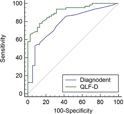
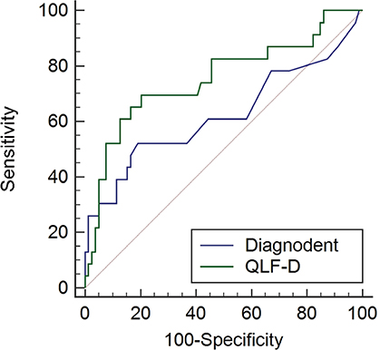
Each method showed an excellent reliability. The correlation coefficient between bitewing radiography and QLF-D, DIAGNOdent were −0.644 and 0.448, respectively, while the value between QLF-D and DIAGNOdent was −0.382. The kappa statistics for bitewing radiography and QLF-D had a higher diagnosis consensus than those for bitewing radiography and DIAGNOdent. The QLF-D was moderately to highly accurate (AUC = 0.753 - 0.908), while DIAGNOdent was moderately to less accurate (AUC = 0.622 - 0.784). All detection methods showed statistically significant correlation and high correlation between the bitewing radiography and QLF-D.
CONCLUSION
QLF-D was found to be a valid and reliable alternative diagnostic method to digital bitewing radiography for in vitro detection of proximal caries.
Keyword
MeSH Terms
Figure
Reference
-
1. Matalon S, Feuerstein O, Kaffe I. Diagnosis of approximal caries: bite-wing radiology versus the Ultrasound Caries Detector. An in vitro study. Oral Surg Oral Med Oral Pathol Oral Radiol Endod. 2003; 95:626–631.2. Murray JJ, Majid ZA. The prevalence and progression of approximal caries in the deciduous dentition in British children. Br Dent J. 1978; 145:161–164.3. Arnold WH, Gaengler P, Kalkutschke L. Three-dimensional reconstruction of approximal subsurface caries lesions in deciduous molars. Clin Oral Investig. 1998; 2:174–179.4. Rayner JA, Southam JC. Pulp changes in deciduous teeth associated with deep carious dentine. J Dent. 1979; 7:39–42.5. Choi SC. About the effect of Bite-wing radiograph’s diagnosis. J Korean Dent Assoc. 1989; 27:688–695.6. White SC, Pharoah MJ. Oral radiology: principles and interpretation. 4th ed. St. Louis: Mosby;2000. p. 272–279.7. Kim BI. QLF Concept and Clinical Imprementation. J Korean Dent Assoc. 2011; 49:443–450.8. Kang SM, Min JH, Kim HN, Kim BI, Kim JB, Jeong SH. In vitro quantification of occlusal caries lesion using QLF-D, ICDAS, DIAGNOdent. J Korean Acad Oral Health. 2014; 38:105–110.9. Pretty IA. Caries detection and diagnosis: novel technologies. J Dent. 2006; 34:727–739.10. Hibst R, Paulus R. Caries detection by red excited fluorescence: investigations on fluorophores. Caries Res. 1999; 33:295.11. Yang J, Dutra V. Utility of radiology, laser fluorescence, and transillumination. Dent Clin North Am. 2005; 49:739–752. vi12. Chong MJ, Seow WK, Purdie DM, Cheng E, Wan V. Visualtactile examination compared with conventional radiography, digital radiography, and Diagnodent in the diagnosis of occlusal occult caries in extracted premolars. Pediatr Dent. 2003; 25:341–349.13. Mepparambath R, S Bhat S, K Hegde S, Anjana G, Sunil M, Mathew S. Comparison of proximal caries detection in primary teeth between laser fluorescence and bitewing radiography: An in vivo study. Int J Clin Pediatr Dent. 2014; 7:163–167.14. Virajsilp V, Thearmontree A, Aryatawong S, Paiboonwarachat D. Comparison of proximal caries detection in primary teeth between laser fluorescence and bitewing radiography. Pediatr Dent. 2005; 27:493–499.15. Shi XQ, Tranaeus S, Angmar-Månsson B. Comparison of QLF and DIAGNOdent for quantification of smooth surface caries. Caries Res. 2001; 35:21–26.16. Farah RA, Drummond BK, Swain MV, Williams S. Relationship between laser fluorescence and enamel hypomineralisation. J Dent. 2008; 36:915–921.17. Kim SH, Lee KH, Kim DE, Park JS. Caries diagnosis by diagnodent›s laser fluorescence detection in vitro. J Korean Acad Pediatr Dent. 2000; 27:24–31.18. Ko HY, Kang SM, Kim HE, Kwon HK, Kim BI. Validation of quantitative light-induced fluorescence-digital (QLF-D) for the detection of approximal caries in vitro. J Dent. 2015; 43:568–575.19. Lussi A, Hack A, Hug I, Heckenberger H, Megert B, Stich H. Detection of approximal caries with a new laser fluorescence device. Caries Res. 2006; 40:97–103.20. Bussaneli DG, Restrepo M, Boldieri T, Albertoni TH, Santos-Pinto L, Cordeiro RC. Proximal caries lesion detection in primary teeth: does this justify the association of diagnostic methods? Lasers Med Sci. 2015; 30:2239–2244.21. Allen MJ, Yen WM. Introduction to measurement theory. Belmont, CA: Wadsworth;1979. p. 76–77.22. Pitts NB. Systems for grading approximal carious lesions and overlaps diagnosed from bitewing radiographs. Proposals for future standardization. Community Dent Oral Epidemiol. 1984; 12:114–122.23. Mejàre I, Gröndahl HG, Carlstedt K, Grever AC, Ottosson E. Accuracy at radiography and probing for the diagnosis of proximal caries. Scand J Dent Res. 1985; 93:178–184.24. Eli I, Weiss EI, Tzohar A, Littner MM, Gelernter I, Kaffe I. Interpretation of bitewing radiographs. Part 1. Evaluation of the presence of approximal lesions. J Dent. 1996; 24:379–383.25. van der Veen MH, de Josselin de Jong E. Application of quantitative light-induced fluorescence for assessing early caries lesions. Monogr Oral Sci. 2000; 17:144–162.26. Buchalla W, Lennon AM, van der Veen MH, Stookey GK. Optimal camera and illumination angulations for detection of interproximal caries using quantitative light-induced fluorescence. Caries Res. 2002; 36:320–326.27. Alwas-Danowska HM, Plasschaert AJ, Suliborski S, Verdonschot EH. Reliability and validity issues of laser fluorescence measurements in occlusal caries diagnosis. J Dent. 2002; 30:129–134.28. Anttonen V, Seppä L, Hausen H. A follow-up study of the use of DIAGNOdent for monitoring fissure caries in children. Community Dent Oral Epidemiol. 2004; 32:312–318.
- Full Text Links
- Actions
-
Cited
- CITED
-
- Close
- Share
- Similar articles
-
- Evaluation of Detection Ability of a Quantitative Light-Induced Fluorescence Digital Device for Initial Secondary Caries Lesion
- Comparison of Diagnostic Validity between Laser Fluorescence Devices in Proximal Caries
- Detecting of Proximal Caries in Primary Molars using Pen-type QLF Device
- Assessment of the Caries Detection Ability of Quantitative Light-induced Fluorescence (QLF) in Primary Teeth
in vitro - Diagnostic Utilization of Laser Fluorescence for Resin Infiltration in Primary Teeth


