Perianal Paget disease: a report of 2 cases
- Affiliations
-
- 1Division of Colorectal Surgery, Department of Surgery, Colorectal Cancer Clinic, Severance Hospital, Yonsei University College of Medicine, Seoul, Korea. namkyuk@yuhs.ac
- 2Department of Pathology, Severance Hospital, Yonsei University College of Medicine, Seoul, Korea.
- 3Department of Plastic and Reconstructive Surgery, Severance Hospital, Yonsei University College of Medicine, Seoul, Korea.
- 4Department of Dermatology and Cutaneous Biology Research Institute, Severance Hospital, Yonsei University College of Medicine, Seoul, Korea.
- KMID: 2396876
- DOI: http://doi.org/10.4174/astr.2017.93.6.336
Abstract
- Extramammary Paget disease (EMPD) is a rare cutaneous neoplasm. Perianal Paget disease (PPD) is a subset of EMPD manifesting perianal lesions. Two cases of PPD in Severance Hospital are described in this article. A 65-year-old female and 78-year-old male patients visited our institution because of an unhealed perianal skin lesion despite treatment for a long period with topical agents. PPD was diagnosed by skin biopsies in both cases, and the patients underwent surgical treatment. Clinical manifestations, preoperative work-ups, and surgical treatments including different reconstruction methods are described in detail. As only sporadic PPD cases have been reported and no standard treatment has been established, we hope that our experience could contribute to improving the diagnosis and treatment of PPD patients.
Keyword
Figure
-
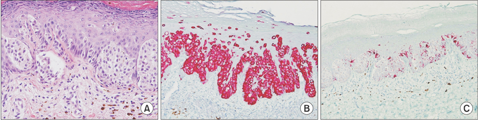
Fig. 1 H&E and IHC stain in case 1. (A) Clusters of large epithelioid cells with pleomorphic nuclei and pale cytoplasm are present in the epidermis, consistent with EMPD (H&E, ×200). (B) Tumor cells show strong immunoreactivity for CK7 with cytoplasmic staining pattern (×100). (C) Tumor cells are negative for HMB-45 IHC, ruling out the possibility of melanoma (×100). CK7, cytokeratin 7; EMPD, extramammary Paget's disease; H&E, hematoxylin and eosin; HMB-45, human melanoma black-45; IHC, immunohistochemistry.

Fig. 2 Case 1. (A) Erythematous perianal skin lesion with slightly elevated patches, erosion, and brownish pigmentation; scout line determined by mapping biopsies (purple). (B) Wide excision with sphincter preservation. (C) Surgical specimen. (D, E) Internal pudendal artery perforator (IPAP) flap. (F) After completion of the IPAP flap and V-Y advancement flap surgery.
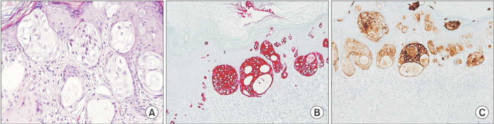
Fig. 3 H&E and IHC in case 2. (A) Clusters of large epithelioid cells with pleomorphic nuclei and pale cytoplasm in the epidermis (H&E, ×200). (B) Tumor cells showing strong immunoreactivity for CK7 (×100; red, in cytoplasm). (C) Tumor cells showing immunoreactivity for CEA (×100; brown, in cytoplasm). CEA, carcinoembryonic antigen; CK7, cytokeratin 7; H&E, hematoxylin and eosin; IHC, immunohistochemistry.
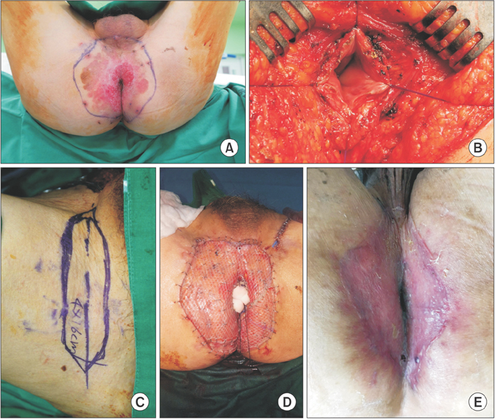
Fig. 4 Case 2. (A) Eroded macules with some whitish patches in the perianal area; scout line determined by mapping biopsies (purple). (B) Wide excision with sphincter preservation. (C, D) Skin graft surgery. (E) Healed wound.
Reference
-
1. Perez DR, Trakarnsanga A, Shia J, Nash GM, Temple LK, Paty PB, et al. Management and outcome of perianal Paget's disease: a 6-decade institutional experience. Dis Colon Rectum. 2014; 57:747–751.2. Minicozzi A, Borzellino G, Momo R, Steccanella F, Pitoni F, de Manzoni G. Perianal Paget's disease: presentation of six cases and literature review. Int J Colorectal Dis. 2010; 25:1–7.3. Herrel LA, Weiss AD, Goodman M, Johnson TV, Osunkoya AO, Delman KA, et al. Extramammary Paget's disease in males: survival outcomes in 495 patients. Ann Surg Oncol. 2015; 22:1625–1630.4. Moller MG, Lugo-Baruqui JA, Milikowski C, Salgado CJ. Staged marginal contoured and central excision technique in the surgical management of perianal Paget's disease. Am J Surg. 2014; 207:485–492.5. Nowak MA, Guerriere-Kovach P, Pathan A, Campbell TE, Deppisch LM. Perianal Paget's disease: distinguishing primary and secondary lesions using immunohistochemical studies including gross cystic disease fluid protein-15 and cytokeratin 20 expression. Arch Pathol Lab Med. 1998; 122:1077–1081.6. Miyamoto A, Akasaka K, Oikawa H, Akasaka T, Masuda T, Maesawa C. Immunohistochemical study of HER2 and TUBB3 proteins in extramammary Paget disease. Am J Dermatopathol. 2010; 32:578–585.7. Sasaki K, Nozaki M, Kikutchi Y, Yamaki T, Soejima K. Reconstruction of perianal skin defect using a V-Y advancement of bilateral gluteus maximus musculocutaneous flaps: reconstruction considering anal cleft and anal function. Br J Plast Surg. 1999; 52:471–475.8. Mizuno H, Iwasaki Y, Noda H, Kawai M, Yamamoto S, Takesue Y. The surgical treatment of perianal paget's disease. Nishinihon J Dermatol. 2001; 63:309–313.9. Brown RS, Lankester KJ, McCormack M, Power DA, Spittle MF. Radiotherapy for perianal Paget's disease. Clin Oncol (R Coll Radiol). 2002; 14:272–284.10. Seckel BR, Schoetz DJ Jr, Coller JA. Skin grafts for circumferential coverage of perianal wounds. Surg Clin North Am. 1985; 65:365–371.
- Full Text Links
-
- Actions
-
Cited
- CITED
-
- Close
- Share
-
- Similar articles
-
- Extramammary Paget's Disease Presenting as a Huge Mass on the Perianal Area
- Extramammary Paget's Disease of the Penis and Scrotum: Report of 2 Cases
- A Case of Extramammary Paget's Disease of the Axilla
- Perianal Paget's Disease Reconstruction with Bilateral Pacman Flap
- Paget's Disease on Penis and Scrotum: A Study of 3 Cases
