Korean J Ophthalmol.
2017 Dec;31(6):548-556. 10.3341/kjo.2016.0118.
Analysis of the Retinal Nerve Fiber Layer Thickness in Alzheimer Disease and Mild Cognitive Impairment
- Affiliations
-
- 1Department of Ophthalmology, Seonam University Myongji Hospital, Goyang, Korea. Kimdk89@empas.com
- KMID: 2396706
- DOI: http://doi.org/10.3341/kjo.2016.0118
Abstract
- PURPOSE
To compare the retinal nerve fiber layer (RNFL) as well as the macula volume and thickness in the eyes of age-matched healthy controls with no cognitive disabilities with those of elderly people with mild cognitive impairment (MCI) or Alzheimer disease (AD). We used optical coherence tomography (OCT) to determine the effectiveness of the above quantities for early diagnosis of MCI or AD.
METHODS
Ninety eyes were considered in this study, split between 30 normal eyes, 30 eyes from patients with MCI, and 30eyes from patients with AD. All subjects underwent ophthalmologic and cognitive examinations, and measurements of the RNFL thickness as well as macular volume and thickness were taken for all patients using OCT.
RESULTS
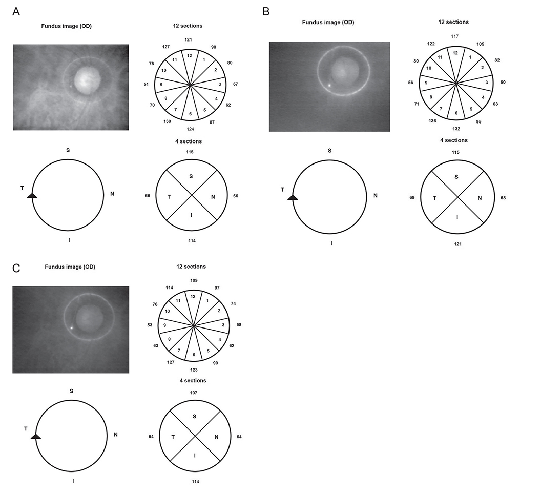
The mean RNFL thickness upon OCT was significantly thinner in the AD group than in the MCI group (p = 0.01). The RNFL was thinner in the superior quadrant in patients with AD when compared to the healthy controls (p = 0.03). The RNFL thicknesses in the inferior, nasal, and temporal quadrants did not differ significantly between the groups. Measurements in the 12 clock-hour zones revealed that zone 11 had a significantly thinner RNFL in the AD group as compared with the healthy control group (p = 0.02). In zone 2, the MCI group had a significantly thinner RNFL than the AD group (p = 0.03).
CONCLUSIONS
Our OCT findings revealed a neuroanatomic difference in the RNFL thickness among the three groups, i.e., the AD, MCI, and healthy control groups. This suggests that a change in average RNFL thickness could be a meaningful index for diagnosing early AD.
Keyword
MeSH Terms
Figure
Reference
-
1. Fratiglioni L, De Ronchi D, Aguero-Torres H. Worldwide prevalence and incidence of dementia. Drugs Aging. 1999; 15:365–375.2. Gao L, Liu Y, Li X, et al. Abnormal retinal nerve fiber layer thickness and macula lutea in patients with mild cognitive impairment and Alzheimer's disease. Arch Gerontol Geriatr. 2015; 60:162–167.3. Braak H, Braak E. Neuropathological stageing of Alzheimer-related changes. Acta Neuropathol. 1991; 82:239–259.4. Leuba G, Saini K. Pathology of subcortical visual centres in relation to cortical degeneration in Alzheimer's disease. Neuropathol Appl Neurobiol. 1995; 21:410–422.5. Hof PR, Vogt BA, Bouras C, Morrison JH. Atypical form of Alzheimer's disease with prominent posterior cortical atrophy: a review of lesion distribution and circuit disconnection in cortical visual pathways. Vision Res. 1997; 37:3609–3625.6. Blanks JC, Torigoe Y, Hinton DR, Blanks RH. Retinal pathology in Alzheimer's disease. I. Ganglion cell loss in foveal/parafoveal retina. Neurobiol Aging. 1996; 17:377–384.7. Trick GL, Trick LR, Morris P, Wolf M. Visual field loss in senile dementia of the Alzheimer's type. Neurology. 1995; 45:68–74.8. Petersen RC, Smith GE, Waring SC, et al. Mild cognitive impairment: clinical characterization and outcome. Arch Neurol. 1999; 56:303–308.9. Kelley BJ, Petersen RC. Alzheimer's disease and mild cognitive impairment. Neurol Clin. 2007; 25:577–609.10. Levey A, Lah J, Goldstein F, et al. Mild cognitive impairment: an opportunity to identify patients at high risk for progression to Alzheimer's disease. Clin Ther. 2006; 28:991–1001.11. Werner P, Korczyn AD. Mild cognitive impairment: conceptual, assessment, ethical, and social issues. Clin Interv Aging. 2008; 3:413–420.12. Petersen RC, Stevens JC, Ganguli M, et al. Practice parameter: early detection of dementia: mild cognitive impairment (an evidence-based review). Report of the Quality Standards Subcommittee of the American Academy of Neurology. Neurology. 2001; 56:1133–1142.13. Winblad B, Palmer K, Kivipelto M, et al. Mild cognitive impairment: beyond controversies, towards a consensus: report of the International Working Group on Mild Cognitive Impairment. J Intern Med. 2004; 256:240–246.14. Petersen RC, Roberts RO, Knopman DS, et al. Mild cognitive impairment: ten years later. Arch Neurol. 2009; 66:1447–1455.15. Morris JC, Cummings J. Mild cognitive impairment (MCI) represents early-stage Alzheimer's disease. J Alzheimers Dis. 2005; 7:235–239.16. Markesbery WR, Schmitt FA, Kryscio RJ, et al. Neuropathologic substrate of mild cognitive impairment. Arch Neurol. 2006; 63:38–46.17. Ikram MK, Cheung CY, Wong TY, Chen CP. Retinal pathology as biomarker for cognitive impairment and Alzheimer's disease. J Neurol Neurosurg Psychiatry. 2012; 83:917–922.18. Hinton DR, Sadun AA, Blanks JC, Miller CA. Optic-nerve degeneration in Alzheimer's disease. N Engl J Med. 1986; 315:485–487.19. Jaffe GJ, Caprioli J. Optical coherence tomography to detect and manage retinal disease and glaucoma. Am J Ophthalmol. 2004; 137:156–169.20. Hassenstein A, Spital G, Scholz F, et al. Optical coherence tomography for macula diagnostics. Review of methods and standardized application concentrating on diagnostic and therapy control of age-related macula degeneration. Ophthalmologe. 2009; 106:116–126.21. Ratchford JN, Quigg ME, Conger A, et al. Optical coherence tomography helps differentiate neuromyelitis optica and MS optic neuropathies. Neurology. 2009; 73:302–308.22. Hajee ME, March WF, Lazzaro DR, et al. Inner retinal layer thinning in Parkinson disease. Arch Ophthalmol. 2009; 127:737–741.23. Ascaso FJ, Cabezon L, Quintanilla MA, et al. Retinal nerve fiber layer thickness measured by optical coherence tomography in patients with schizophrenia: a short report. Eur J Psychiatry. 2010; 24:227–235.24. Parisi V, Restuccia R, Fattapposta F, et al. Morphological and functional retinal impairment in Alzheimer's disease patients. Clin Neurophysiol. 2001; 112:1860–1867.25. Iseri PK, Altinas O, Tokay T, Yuksel N. Relationship between cognitive impairment and retinal morphological and visual functional abnormalities in Alzheimer disease. J Neuroophthalmol. 2006; 26:18–24.26. Berisha F, Feke GT, Trempe CL, et al. Retinal abnormalities in early Alzheimer's disease. Invest Ophthalmol Vis Sci. 2007; 48:2285–2289.27. Paquet C, Boissonnot M, Roger F, et al. Abnormal retinal thickness in patients with mild cognitive impairment and Alzheimer's disease. Neurosci Lett. 2007; 420:97–99.28. Kesler A, Vakhapova V, Korczyn AD, et al. Retinal thickness in patients with mild cognitive impairment and Alzheimer's disease. Clin Neurol Neurosurg. 2011; 113:523–526.29. Cheung CY, Ong YT, Hilal S, et al. Retinal ganglion cell analysis using high-definition optical coherence tomography in patients with mild cognitive impairment and Alzheimer's disease. J Alzheimers Dis. 2015; 45:45–56.30. Kirbas S, Turkyilmaz K, Anlar O, et al. Retinal nerve fiber layer thickness in patients with Alzheimer disease. J Neuroophthalmol. 2013; 33:58–61.31. Liu D, Zhang L, Li Z, et al. Thinner changes of the retinal nerve fiber layer in patients with mild cognitive impairment and Alzheimer's disease. BMC Neurol. 2015; 15:14.32. Blanks JC, Schmidt SY, Torigoe Y, et al. Retinal pathology in Alzheimer's disease. II. Regional neuron loss and glial changes in GCL. Neurobiol Aging. 1996; 17:385–395.33. Lamirel C, Newman N, Biousse V. The use of optical coherence tomography in neurology. Rev Neurol Dis. 2009; 6:E105–E120.34. Sakata LM, Deleon-Ortega J, Sakata V, Girkin CA. Optical coherence tomography of the retina and optic nerve: a review. Clin Exp Ophthalmol. 2009; 37:90–99.35. Portet F, Ousset PJ, Visser PJ, et al. Mild cognitive impairment (MCI) in medical practice: a critical review of the concept and new diagnostic procedure. Report of the MCI Working Group of the European Consortium on Alzheimer's Disease. J Neurol Neurosurg Psychiatry. 2006; 77:714–718.36. American Psychiatric Association. Diagnostic and statistical manual of mental disorders (IV-TR). 4th ed. Washington, DC: American Psychiatric Association;2000. p. 413–435.37. Blennow K, Hampel H. CSF markers for incipient Alzheimer's disease. Lancet Neurol. 2003; 2:605–613.38. Galasko D. Biomarkers for Alzheimer's disease: clinical needs and application. J Alzheimers Dis. 2005; 8:339–346.39. He XF, Liu YT, Peng C, et al. Optical coherence tomography assessed retinal nerve fiber layer thickness in patients with Alzheimer's disease: a meta-analysis. Int J Ophthalmol. 2012; 5:401–405.40. Frohman EM, Fujimoto JG, Frohman TC, et al. Optical coherence tomography: a window into the mechanisms of multiple sclerosis. Nat Clin Pract Neurol. 2008; 4:664–675.41. Chi Y, Wang YH, Yang L. The investigation of retinal nerve fiber loss in Alzheimer's disease. Zhonghua Yan Ke Za Zhi. 2010; 46:134–139.42. Schmidtke K, Hermeneit S. High rate of conversion to Alzheimer's disease in a cohort of amnestic MCI patients. Int Psychogeriatr. 2008; 20:96–108.43. Ascaso FJ, Cruz N, Modrego PJ, et al. Retinal alterations in mild cognitive impairment and Alzheimer's disease: an optical coherence tomography study. J Neurol. 2014; 261:1522–1530.44. Feuer WJ, Budenz DL, Anderson DR, et al. Topographic differences in the age-related changes in the retinal nerve fiber layer of normal eyes measured by Stratus optical coherence tomography. J Glaucoma. 2011; 20:133–138.45. Shi Z, Wu Y, Wang M, et al. Greater attenuation of retinal nerve fiber layer thickness in Alzheimer's disease patients. J Alzheimers Dis. 2014; 40:277–283.46. Shen Y, Liu L, Cheng Y, et al. Retinal nerve fiber layer thickness is associated with episodic memory deficit in mild cognitive impairment patients. Curr Alzheimer Res. 2014; 11:259–266.
- Full Text Links
- Actions
-
Cited
- CITED
-
- Close
- Share
- Similar articles
-
- Reproducibility of Retinal Nerve Fiber Layer Thickness Evaluation by Nerve Fiber Analyzer
- Biometry of Retinal Nerve Fiber Layer Thickness by NFA
- Decreased Retinal Thickness in Patients With Alzheimer's Disease
- Associations of Peripapillary Retinal Nerve Fiber Layer and Macular Retinal Layer Thickness with Serum Homocysteine Concentration
- Influence of Diabetes Mellitus on the Retinal Ne rve Fiber Layer Thickness Measurement by Nerve Fiber Analyzer


