Ann Dermatol.
2017 Dec;29(6):809-810. 10.5021/ad.2017.29.6.809.
Primary Angiosarcoma of the Skin Presenting as Mild Erythema
- Affiliations
-
- 1Department of Dermatology, Kyungpook National University School of Medicine, Daegu, Korea. weonju@knu.ac.kr
- KMID: 2395198
- DOI: http://doi.org/10.5021/ad.2017.29.6.809
Abstract
- No abstract available.
MeSH Terms
Figure
-
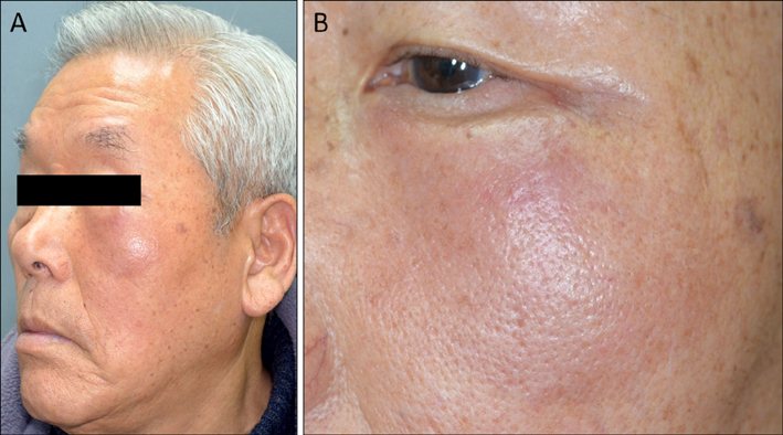
Fig. 1 (A, B) Ill-demarcated, indurated erythema in the left zygomatic area.

Fig. 2 An irregular anastomosing vascular space lined with atypical endothelial cells (H&E; A: ×40, B: ×100, C: ×200). Immunohistochemical analysis of the lesion shows tumor cells stained for (D) CD31, (E) D2-40, or (F) Prox-1. D~F: ×100.
Reference
-
1. Guadagnolo BA, Zagars GK, Araujo D, Ravi V, Shellenberger TD, Sturgis EM. Outcomes after definitive treatment for cutaneous angiosarcoma of the face and scalp. Head Neck. 2011; 33:661–667.
Article2. Kim JE, Kim BJ, Kang H. A recurrent angiosarcoma isolated to the eyelid without the recurrence on the primary lesion of the forehead. Ann Dermatol. 2014; 26:231–235.
Article3. Lemanski N, Farber M, Carruth BP, Wladis EJ. Primary adnexal angiosarcoma masquerading as periorbital hematoma. Surv Ophthalmol. 2014; 59:655–659.4. Mentzel T, Kutzner H, Wollina U. Cutaneous angiosarcoma of the face: clinicopathologic and immunohistochemical study of a case resembling rosacea clinically. J Am Acad Dermatol. 1998; 38:837–840.
Article5. Aguila LI, Sánchez JL. Angiosarcoma of the face resembling rhinophyma. J Am Acad Dermatol. 2003; 49:530–531.
Article
