Ann Dermatol.
2017 Apr;29(2):229-232. 10.5021/ad.2017.29.2.229.
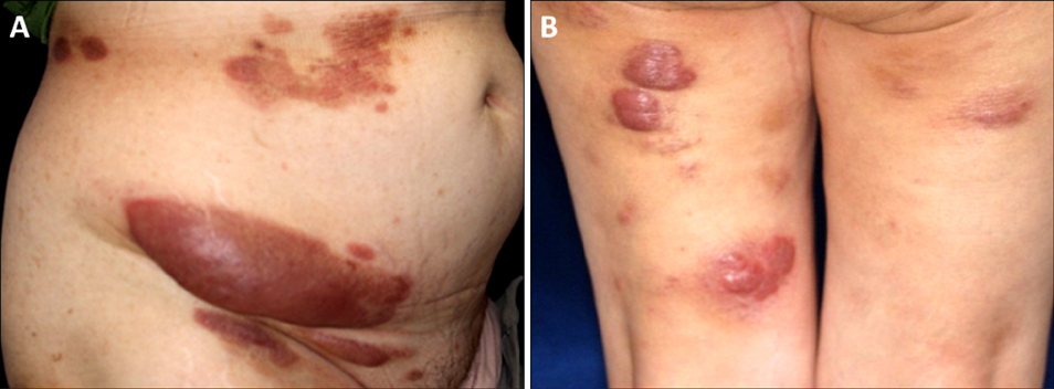
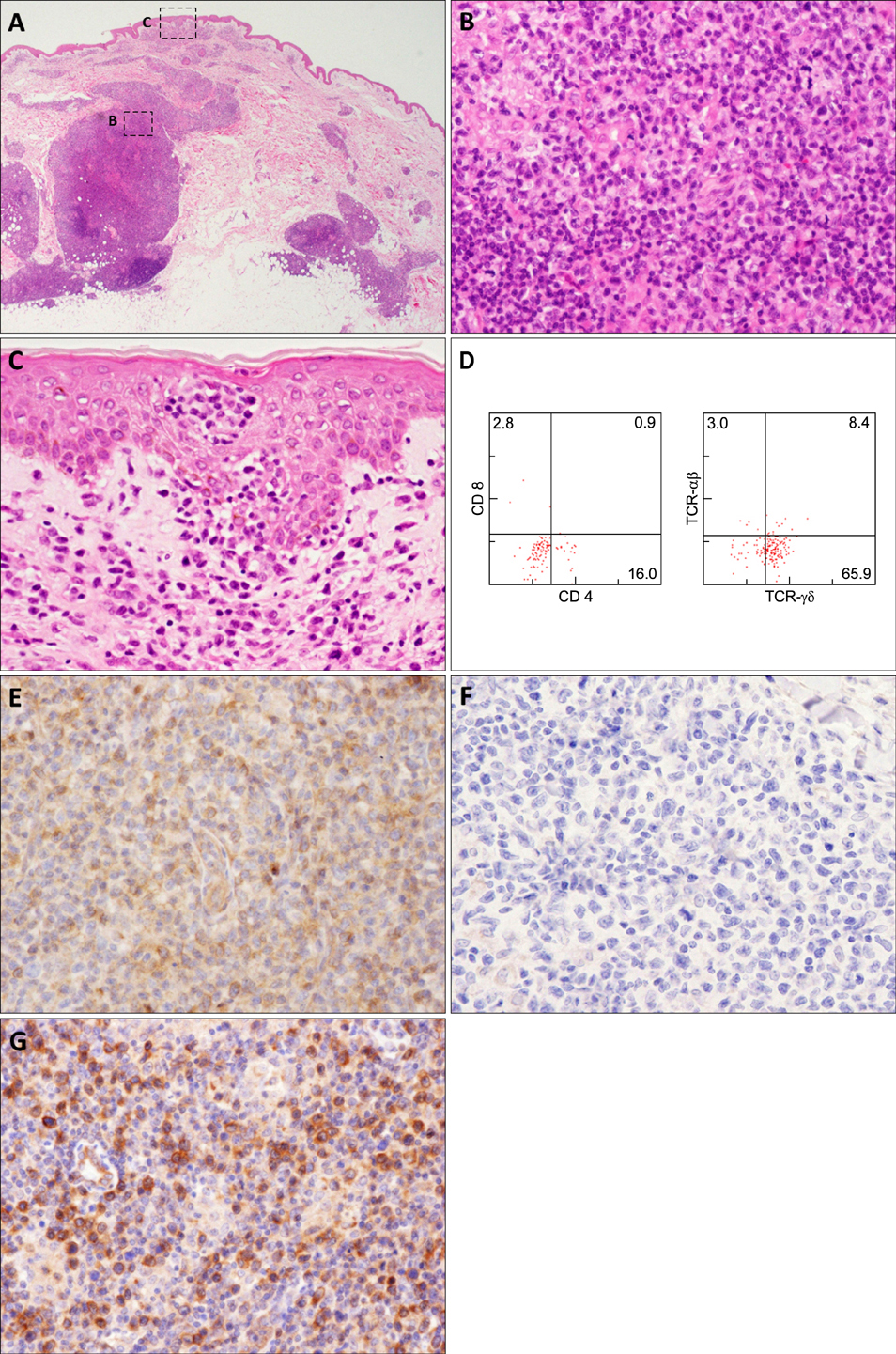
A Case of Primary Cutaneous Gamma-Delta T-Cell Lymphoma with Pautrier Microabscess
- Affiliations
-
- 1Department of Dermatology, Graduate School of Medical and Dental Sciences, Tokyo Medical and Dental University, Tokyo, Japan. tnamderm@tmd.ac.jp
- 2Department of Pathology, Graduate School of Medical and Dental Sciences, Tokyo Medical and Dental University, Tokyo, Japan.
- KMID: 2394851
- DOI: http://doi.org/10.5021/ad.2017.29.2.229
Abstract
- No abstract available.
MeSH Terms
Figure
Reference
-
1. Toro JR, Liewehr DJ, Pabby N, Sorbara L, Raffeld M, Steinberg SM, et al. Gamma-delta T-cell phenotype is associated with significantly decreased survival in cutaneous T-cell lymphoma. Blood. 2003; 101:3407–3412.
Article2. Tripodo C, Iannitto E, Florena AM, Pucillo CE, Piccaluga PP, Franco V, et al. Gamma-delta T-cell lymphomas. Nat Rev Clin Oncol. 2009; 6:707–717.
Article3. Rodríguez-Pinilla SM, Ortiz-Romero PL, Monsalvez V, Tomás IE, Almagro M, Sevilla A, et al. TCR-γ expression in primary cutaneous T-cell lymphomas. Am J Surg Pathol. 2013; 37:375–384.
Article4. Guitart J, Weisenburger DD, Subtil A, Kim E, Wood G, Duvic M, et al. Cutaneous γδ T-cell lymphomas: a spectrum of presentations with overlap with other cytotoxic lymphomas. Am J Surg Pathol. 2012; 36:1656–1665.5. Suga H, Sugaya M, Miyagaki T, Ohmatsu H, Fujita H, Sato S. Primary cutaneous γδ T-cell lymphoma following mycosis fungoides. Int J Dermatol. 2014; 53:e82–e84.
Article
- Full Text Links
- Actions
-
Cited
- CITED
-
- Close
- Share
- Similar articles
-
- Fatal Cutaneous gamma/delta T-Cell Lymphoma with Central Nerve System Metastasis
- A Case of Extranodall NK/T-cell Lymphoma, Nasal type
- A Case of Primary Cutaneous Marginal Zone B-cell Lymphoma
- A Case of Primary Nasal CD56+ NK/T cell Lymphoma with Cutaneous Involvement
- A Case of Non-T,Non-B Primary Cutaneous Lymphoblastic Lymphoma


