Ann Dermatol.
2017 Apr;29(2):215-218. 10.5021/ad.2017.29.2.215.
Degos-Like Lesions Associated with Systemic Lupus Erythematosus
- Affiliations
-
- 1Department of Dermatology, Kosin University College of Medicine, Busan, Korea. ksderm98@unitel.co.kr
- 2Department of Internal Medicine, Kosin University College of Medicine, Busan, Korea.
- KMID: 2394847
- DOI: http://doi.org/10.5021/ad.2017.29.2.215
Abstract
- Degos disease, also referred to as malignant atrophic papulosis, was first described in 1941 by Köhlmeier and was independently described by Degos in 1942. Degos disease is characterized by diffuse, papular skin eruptions with porcelain-white centers and slightly raised erythematous telangiectatic rims associated with bowel infarction. Although the etiology of Degos disease is unknown, autoimmune diseases, coagulation disorders, and vasculitis have all been considered as underlying pathogenic mechanisms. Approximately 15% of Degos disease have a benign course limited to the skin and no history of gastrointestinal or central nervous system (CNS) involvement. A 29-year-old female with history of systemic lupus erythematosus (SLE) presented with a 2-year history of asymptomatic lesions on the dorsum of all fingers and both knees. The patient had only skin lesions and no gastrointestinal or CNS vasculitis symptoms. Her skin lesions were umbilicated, atrophic porcelain-white lesions with a rim of erythema. On the basis of clinical, histologic, and laboratory findings, a diagnosis of Degos-like lesions associated with SLE was made. The patient had been treated for SLE for 7 years. Her treatment regimen was maintained over a 2 month follow-up period, and the skin lesions improved slightly with no development of new lesions.
MeSH Terms
Figure
-
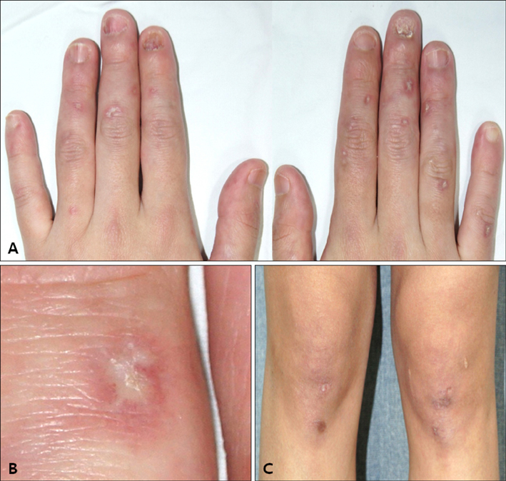
Fig. 1 (A~C) Multiple erythematous papules with porcelain-white scars surrounded by an erythematous rim were seen on dorsum of all fingers and both knees.
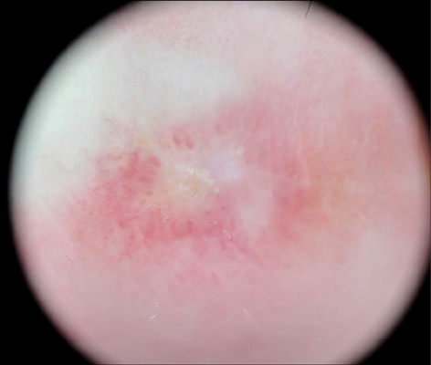
Fig. 2 On dermoscopy, a central, whitish, structureless area surrounded by an erythematous rim is seen (×10).
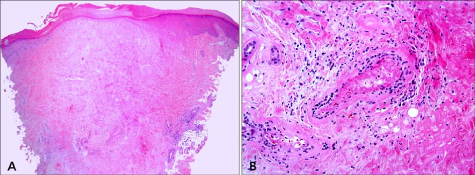
Fig. 3 (A) Histologic findings showed hyperkeratosis, epidermal atrophy, and dermal sclerosis in the central portion (H&E, ×40). (B) Lymphocytic infiltration around vessels, fibrinoid necrosis of the vessel wall, and thrombus deposition in the lumen were seen (H&E, ×400).
Reference
-
1. Köhlmeier W. Multiple hautnekrosen bei thromboangiitis obliterans. Arch Dermatol Syphilol. 1941; 181:792–793.2. Lipsker D. Malignant atrophic papulosis (Degos disease). In : Goldsmith LA, Katz SI, Gilchrest BA, Paller AS, Leffell DJ, Wolff K, editors. Fitzpatrick's dermatology in general medicine. 8th ed. New York: McGraw Hill;2012. p. 2072–2076.3. Lee JH, Ryu HJ, Park YB, Choi BY, Lee EY, Lee YJ, et al. A case of systemic lupus erythematosus with Degos' disease. J Korean Rheum Assoc. 2007; 14:256–262.
Article4. Magro CM, Poe JC, Kim C, Shapiro L, Nuovo G, Crow MK, et al. Degos disease: a C5b-9/interferon-α-mediated endotheliopathy syndrome. Am J Clin Pathol. 2011; 135:599–610.5. Theodoridis A, Makrantonaki E, Zouboulis CC. Malignant atrophic papulosis (Köhlmeier-Degos disease)-a review. Orphanet J Rare Dis. 2013; 8:10.6. Subbiah P, Wijdicks E, Muenter M, Carter J, Connolly S. Skin lesion with a fatal neurologic outcome (Degos' disease). Neurology. 1996; 46:636–640.
Article7. Tsao H, Busam K, Barnhill RL, Haynes HA. Lesions resembling malignant atrophic papulosis in a patient with dermatomyositis. J Am Acad Dermatol. 1997; 36:317–319.
Article8. Ball E, Newburger A, Ackerman AB. Degos' disease: a distinctive pattern of disease, chiefly of lupus erythematosus, and not a specific disease per se. Am J Dermatopathol. 2003; 25:308–320.
Article9. Cardinali C, Caproni M, Bernacchi E, Amato L, Fabbri P. The spectrum of cutaneous manifestations in lupus erythematosus--the Italian experience. Lupus. 2000; 9:417–423.
Article10. Ramos-Casals M, Nardi N, Lagrutta M, Brito-Zerón P, Bové A, Delgado G, et al. Vasculitis in systemic lupus erythematosus: prevalence and clinical characteristics in 670 patients. Medicine (Baltimore). 2006; 85:95–104.11. Anker JP, Kaminska-Winciorek G, Lallas A, Nicoletti S, Januszewski K, Mazzei ME, et al. The dermoscopic variability of Degos disease at different stages of progression. Dermatol Pract Concept. 2014; 4:59–61.
Article12. Harvell JD, Williford PL, White WL. Benign cutaneous Degos' disease: a case report with emphasis on histopathology as papules chronologically evolve. Am J Dermatopathol. 2001; 23:116–123.13. Kim YJ, Yun SJ, Lee SC, Lee JB. Degos disease associated with Behçet's disease. Ann Dermatol. 2015; 27:235–236.
Article14. Melnik B, Hariry H, Vakilzadeh F, Gropp C, Sitzer G. Malignant atrophic papulosis (Köhlmeier-Degos disease). Failure to respond to interferon alpha-2a, pentoxifylline and aspirin. Hautarzt. 2002; 53:618–621.
Article
- Full Text Links
-
- Actions
-
Cited
- CITED
-
- Close
- Share
-
- Similar articles
-
- A Case of Systemic Lupus Erythematosus with Degos' Disease
- Papulosquamous Skin Lesions in Systemic Lupus Erythematosus
- A Case of Transverse Myelitis as a First Manifestation of Systemic Lupus Erythematosus
- Bullous systemic lupus erythematosus presented with localized mechanobullous lesions
- A Case Of Systemic Lupus Erythematosus Associated With Hyperthyroidism And Severe Retinopathy
