Implant impression using closed mouth impression technique: a case report
- Affiliations
-
- 1Dental Clinic Center, Pusan National University Hosptial, Busan, Republic of Korea.
- 2Nanum E Dental clinic, Suwon, Repubilc of Korea.
- 3Department of Prosthodontics, In-Je University Haeundae Paik Hospital, Busan, Republic of Korea.
- 4Department of Prosthodontics, School of Dentistry, Pusan National University, Yangsan, Republic of Korea. p-venus79@hanmail.net
- KMID: 2394646
- DOI: http://doi.org/10.14368/jdras.2017.33.3.223
Abstract
- Closed mouth impression technique by using bite tray is preferred for single tooth impression taking. However, for implant impression taking, open mouth impression technique by using single arch tray is generally used whether it is for single implant or multiple implant. Closed mouth impression technique by using bite tray can save time and materials. It also decreases the chance of error occurrence when a model is mounted on an articulator. In this case report, we tried to show a satisfying result of fabricating single implant fixed prosthodontics after bite tray impression taking by using two different copings for closed mouth impression.
MeSH Terms
Figure
-
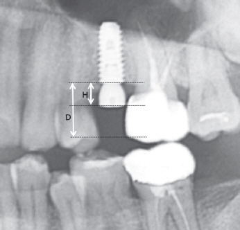
Fig. 1 Measurements performed on radiograph. H is 4 mm. Based on the radiographic view, D is supposed to be more than two times of H. H, height of healing abutment; D, distance from the fixture platform to the expected occlusal surface.

Fig. 2 (A) Impression coping, (B) Impression cap, (C) Impression coping with impression cap. The total length above the fixture platform is 8 mm. a, round nutch; b, flat nutch.
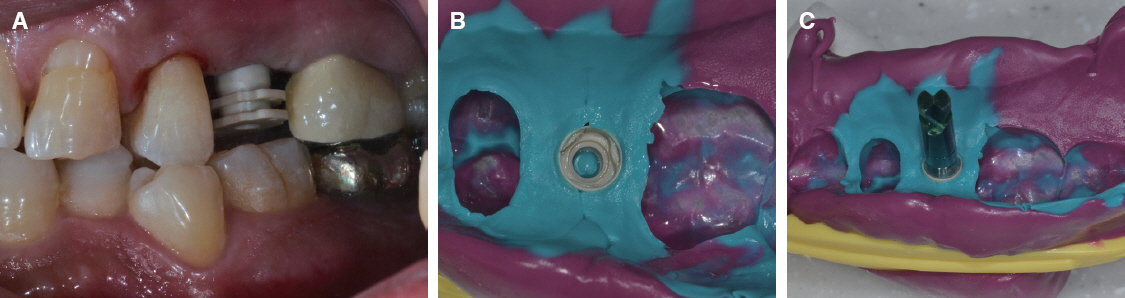
Fig. 3 (A) Impression coping with cap is connected to the implant fixture. (B) Impression cap was removed from the mouth together with the set impression while impression coping was retained in the mouth. (C) Reposition the impression coping connected with the laboratory analog in the impression cap.
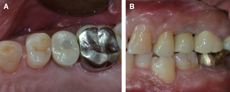
Fig. 4 Six months after definitive prosthesis delivery. (A) Occlusal view. (B) Lateral view.

Fig. 5 (A) Impression coping connected with bite impression coping driver. (B) Intraoral view of impression coping in place. (C) The patient is biting the bite tray according to the conventional impression taking method.

Fig. 6 (A) Impression body was removed from the oral cavity while impression coping remained in the mouth. The shape of the coping in the impression shows parallel two faces (arrows) facing each other. (B) The impression copings removed from the mouth were repositioned on the impression after connecting with the laboratory analog.
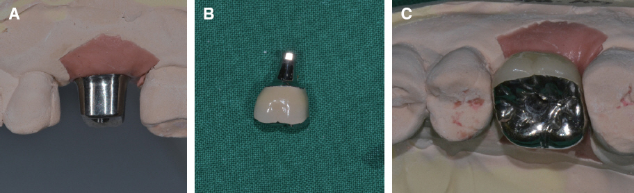
Fig. 7 (A) Customized abutment on working cast. (B) Customized abutment and PFM crown. (C) Restoration on the definitive model.
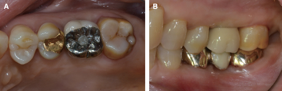
Fig. 8 Five months after definitive prosthesis delivery. (A) Occlusal view. (B) Lateral view.
Reference
-
References
1. de Lima LM, Borges GA, Junior LH, Spohr AM. In vivo study of the accuracy of dual-arch impressions. J Int Oral Health. 2014; 6:50–5. PMID: 25083032. PMCID: PMC4109244.2. Burke FJ, Crisp RJ. A practice-based assessment of the handling of a fast-setting polyvinyl siloxane impression material used with the dual-arch tray technique. Quintessence Int. 2001; 32:805–10. PMID: 11820050.3. Carr AB. Comparison of impression techniques for a five-implant mandibular model. Int J Oral Maxillofac Implants. 1991; 6:448–55. PMID: 1820314.4. Sekine H, Komiyama Y, Potta H, Yoshida K. Mobility characteristics and tactile sensitivity of osseointegrated fixture-supporting systems. van Steenberghe D, Albrektsson T, Brånemark PI, Henry PJ, Holt R, Liden G, editors. Tissue integration in oral and maxillofacial reconstruction. Amsterdam: Excerpta Medica;1986. p. 326–32.5. Tautin FS. Impression making for osseointegrated dentures. J Prosthet Dent. 1985; 54:250–1. DOI: 10.1016/0022-3913(85)90299-9.6. Assif D, Fenton A, Zarb G, Schmitt A. Comparative accuracy of implant impression procedures. Int J Periodontics Restorative Dent. 1992; 12:112–21. DOI: 10.1097/00008505-199200140-00013.7. Parker MH, Cameron SM, Hughbanks JC, Reid DE. Comparison of occlusal contacts in maximum intercuspation for two impression techniques. J Prosthet Dent. 1997; 78:255–9. DOI: 10.1016/S0022-3913(97)70023-4.8. Suzuki Y, Shimpo H, Ohkubo C, Kurtz KS. Fabrication of fixed implant prostheses using function bite impression technique (FBI technique). J Prosthodont Res. 2012; 56:293–6. DOI: 10.1016/j.jpor.2012.02.005. PMID: 22609223.9. Liou AD, Nicholls JI, Yuodelis RA, Brudvik JS. Accuracy of replacing three tapered transfer impression copings in two elastomeric impression materials. Int J Prosthodont. 1993; 6:377–83. PMID: 8240649.10. Siamos G, Winkler S, Boberick KG. Relationship between implant preload and screw loosening on implant-supported prostheses. J Oral Implantol. 2002; 28:67–73. DOI: 10.1563/1548-1336(2002)028<0067:TRBIPA>2.3.CO;2.11. Adell R, Lekholm U, Rockler B, Brånemark PI. A 15-year study of osseointegrated implants in the treatment of the edentulous jaw. Int J Oral Surg. 1981; 10:387–416. DOI: 10.1016/S0300-9785(81)80077-4.12. Chee W, Jivraj S. Impression techniques for implant dentistry. Br Dent J. 2006; 201:429–32. DOI: 10.1038/sj.bdj.4814118. PMID: 17031344.13. Neobiotech. Pickcap Kit. Available from: http://www.neobiotechus.com/index.php/others/pick-cap-kit.html. (updated 2017 July 11).14. Ockert-Eriksson G, Eriksson A, Lockowandt P, Eriksson O. Materials for interocclusal records and their ability to reproduce a 3-dimensional jaw relationship. Int J Prosthodont. 2000; 13:152–8. PMID: 11203625.
- Full Text Links
-
- Actions
-
Cited
- CITED
-
- Close
- Share
-
- Similar articles
-
- Implant overdenture with closed impression technique for patient with Myasthenia Gravis: A case report
- Open and Closed Mouth Impression Techniques for Mandibular Implant Overdenture: Two Cases Report
- Complete denture made with closed-mouth impression technique on severely atrophied edentulous jaw
- Complete denture rehabilitation of edentulous patient with severe alveolar bone resorption andcondyle fracture using gothic arch tracing and closed mouth impression technique: A case report
- Comparison of conventional and suction-effective complete denture in a fully edentulous patient: a case report
