Maxillary removable partial overdenture within Locator Root Attachment in a retained root: a case report
- Affiliations
-
- 1Design Dental Cilinic, Gwangju, Republic of Korea. ddcsjh@gmail.com
- KMID: 2394643
- DOI: http://doi.org/10.14368/jdras.2017.33.3.199
Abstract
- If maintaining oral few retained root when planning the removable prosthesis, it is possible to obtain a more comfortable and functional result. In this case report, "˜Locator Root Attachment' was used to maxillary removable overdenture in patients with a few teeth and retained root. A functionally proper clinical result from attachment after clinical and radiographic analysis was observed in this case.
Keyword
Figure
-
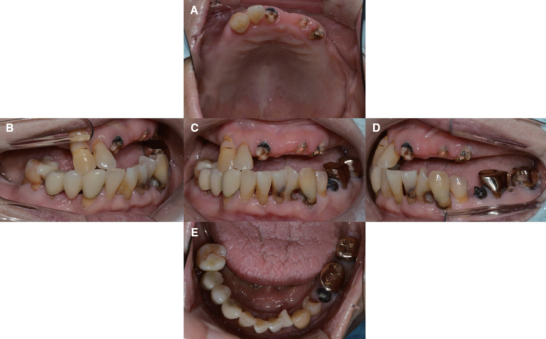
Fig. 1 Preoperative photograph: intraoral view. (A) Occlusal view of maxilla, (B) Lateral view (right side), (C) Frontal view, (D) Lateral view (left side), (E) Occlusal view of mandible.
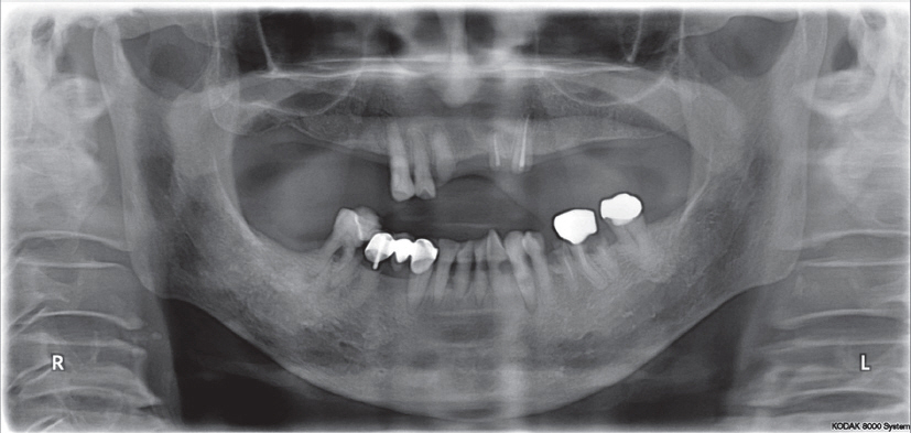
Fig. 2 Preoperative photograph: radiographic view.
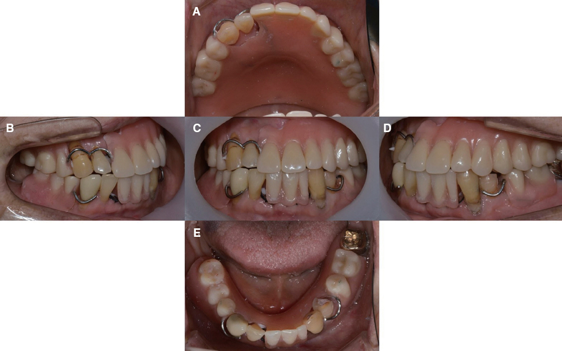
Fig. 3 Interim prostheses (temporary denture). (A) Occlusal view of maxilla, (B) Lateral view (right side), (C) Frontal view, (D) Lateral view (left side), (E) Occlusal view of mandible.
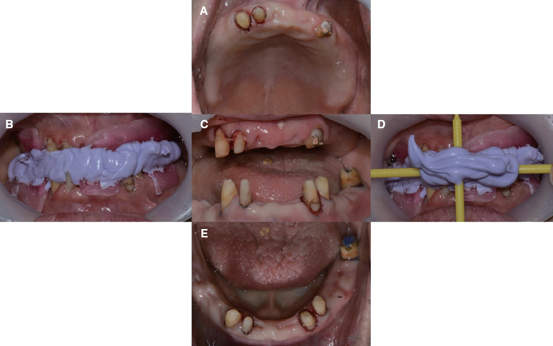
Fig. 4 Preparation photograph. (A) Occlusal view of maxilla, (B) Lateral view (right side), (C) Frontal view, (D) Lateral view (left side), (E) Occlusal view of mandible.
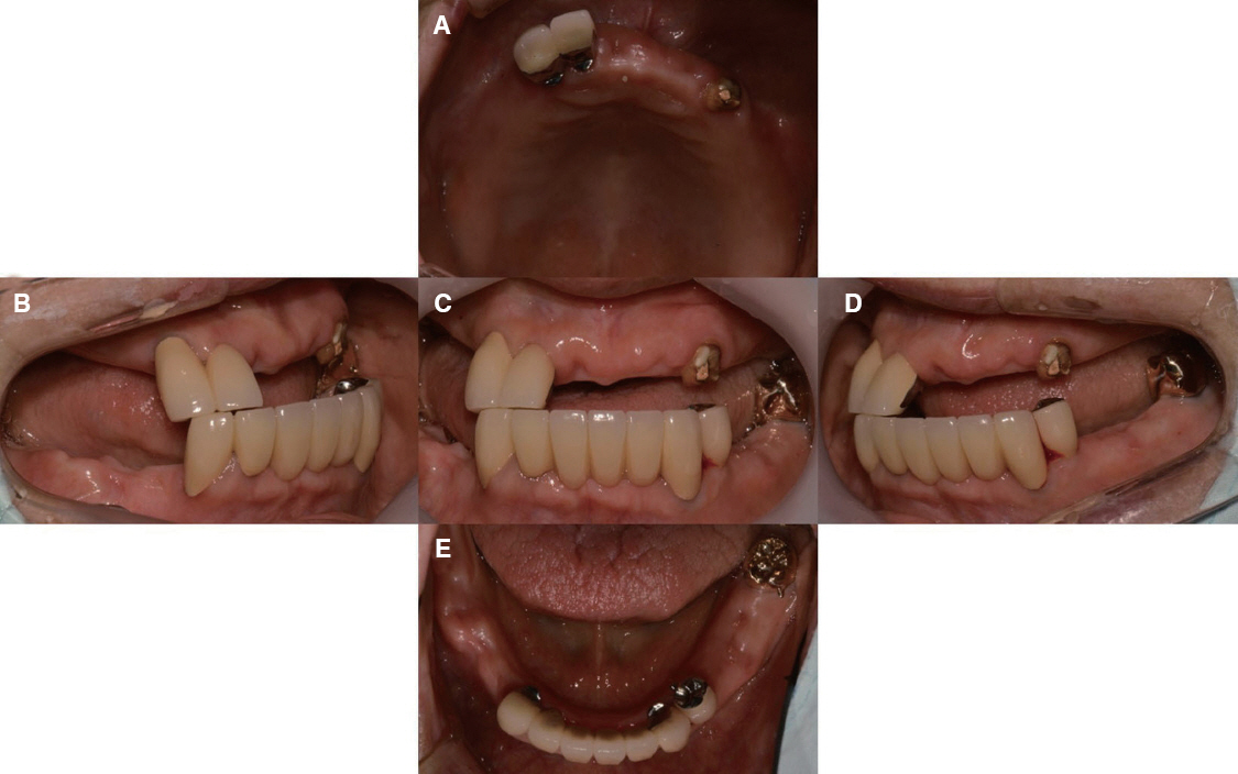
Fig. 5 Definitive restoration (surveyed crown for removable partial denture). (A) Occlusal view of maxilla, (B) Lateral view (right side), (C) Frontal view, (D) Lateral view (left side), (E) Occlusal view of mandible.
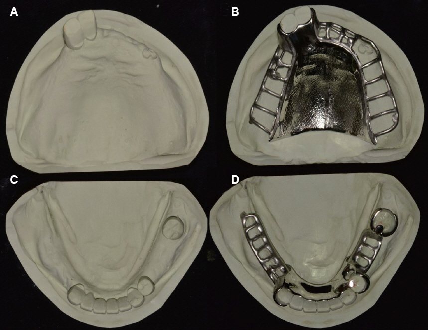
Fig. 6 Master cast and framework. (A) Master cast of maxilla, (B) Framework of maxilla RPD, (C) Master cast of mandible, (D) Framework of mandible RPD.
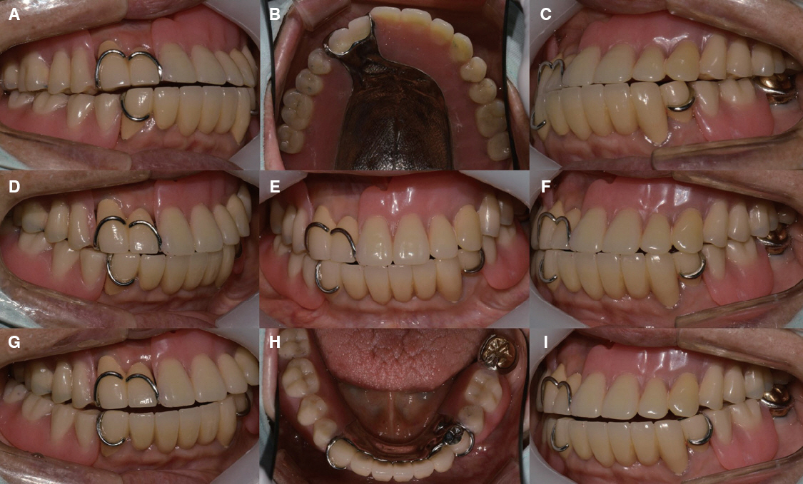
Fig. 7 Definitive restoration (removable partial denture). (A) Non-working side of right lateral view, (B) Occlusal view of maxilla, (C) Non-working side of left lateral view, (D) Lateral view (right side), (E) Frontal view, (F) Lateral view (left side), (G) Working side of right lateral view, (H) Occlusal view of mandible, (I) Working side of left lateral view.
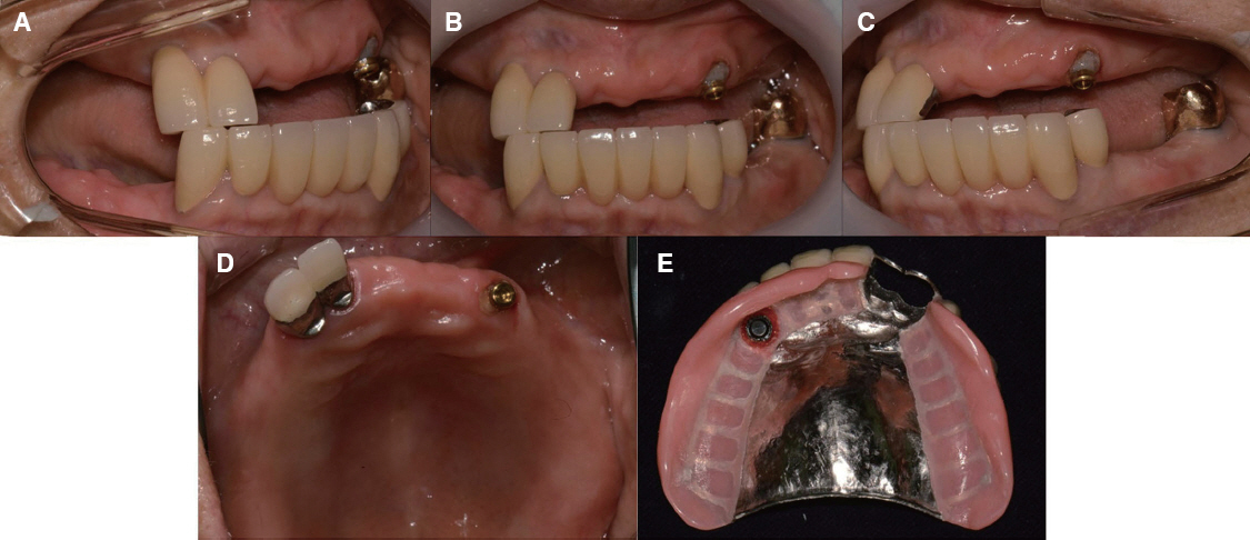
Fig. 8 Locator Root Attachment application. (A) Lateral view (right side), (B) Frontal view, (C) Lateral view (left side), (D) Occlusal view of maxilla, (E) overdenture inner surface.
Reference
-
References
1. Zarb GA, Hobkirk J, Eckert S, Jacob R. Prosthodontic treatment for edentulous patients: complete dentures and implant-supported prostheses. 12th ed. St. Louis: Elsevier Health Sciences;2013. p. 160–76.2. Langer Y, Langer A. Root-retained overdentures: Part I - Biomechanical and clinical aspects. J Prosthet Dent. 1991; 66:784–9. DOI: 10.1016/0022-3913(91)90416-T.3. Renner RP, Gomes BC, Shakun ML, Baer PN, Davis RK, Camp P. Four-year longitudinal study of the periodontal health status of overdenture patients. J Prosthet Dent. 1984; 51:593–8. DOI: 10.1016/0022-3913(84)90399-8.4. Trakas T, Michalakis K, Kang K, Hirayama H. Attachment systems for implant retained overdentures: a literature review. Implant Dent. 2006; 15:24–34. DOI: 10.1097/01.id.0000202419.21665.36. PMID: 16569958.5. Schneider AL, Kurtzman GM. Bar overdentures utilizing the Locator attachment. Gen Dent. 2001; 49:210–4. PMID: 12004703.
- Full Text Links
-
- Actions
-
Cited
- CITED
-
- Close
- Share
-
- Similar articles
-
- A PHOTOELASTIC ANALYSIS ON TOOTH SUPPORTING STRUCTURE AND RESIDUAL RIDGE ACCORDING TO DENTURE DESIGN FOR REMAINING MANDIBULAR CANINES
- The Implant Retained Overdenture by Locator Attachments on the Edentulous Mandible: A Case Report
- Mandibular implant overdenture using Locator R-Tx attachment: A case report
- Removable partial denture restoration using single implant supported with Locator(R) attachment in a crossed occlusion patient: a case report
- Implant-retained overdenture with CM LOC ® Pekkton® in maxillary edentulous patient
