The dimension analysis of prepared natural teeth for developing customized zirconia block
- Affiliations
-
- 1Department of Prosthodontics, Seoul National University School of Dentistry, Seoul, Republic of Korea. proshan@snu.ac.kr
- KMID: 2393909
- DOI: http://doi.org/10.4047/jkap.2017.55.4.381
Abstract
- PURPOSE
Unpredictable shrinkage of zirconia during sintering process causes discrepancy. Therefore, there have been attempts to reduce discrepancy by milling zirconia after sintering. However, due to the hardness of sintered zirconia, milling takes longer time, causes damage to the machine and causes chip formation. With customized zirconia block using the mean dimension of prepared natural dentition, it is expected to overcome these shortcomings.
MATERIALS AND METHODS
The mean dimension of prepared natural dentition was analyzed as STL file after scanning of prepared teeth treated at SNUDH. The transverse, frontal and sagittal planes were set using Mimics and Photoshop. 3D volume was projected on each plane, and the outer line was measured through external tangent line, and the inner line was measured through inflection point of tangent line.
RESULTS
The mean height of prepared incisal (N = 57) is 6.60 ± 1.05 mm, mesiodistal length is 2.98 ± 0.73 mm, buccolingual length is 2.04 ± 0.73 mm. The mean height of prepared premolar (N = 15) is 5.37 ± 1.49 mm, mesiodistal length is 4.10 ± 1.78 mm, buccolingual length is 5.86 ± 1.55 mm. And the mean height of prepared molar (N = 13) is 5.11 ± 1.29 mm, mesiodistal length is 6.80 ± 1.18 mm, buccolingual length is 7.34 ± 1.40 mm.
CONCLUSION
Using the mean dimension of prepared natural dentition, it is expected to be able to fabricate customized zirconia block.
Keyword
Figure
-
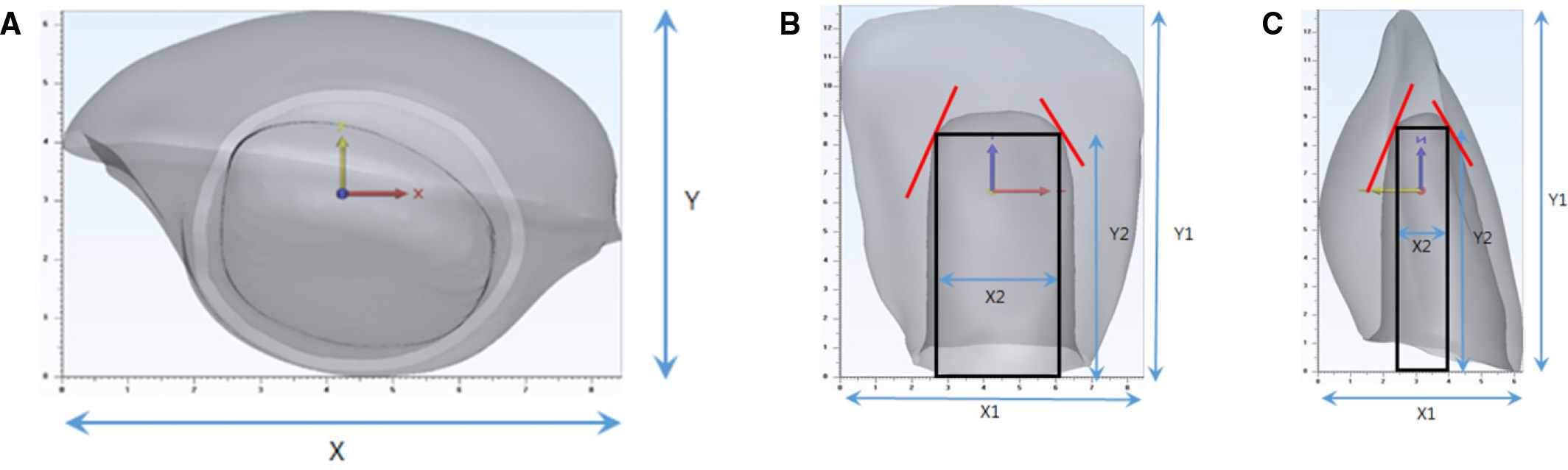
Fig. 1. 3D volume was projected on each plane. The outer line was measured through external tangent line, and inner line was measured through inflection point of tangent line using Mimics auto setup. (A) Transverse Plane, (B) Frontal Plane, (C) Sagittal Plane.
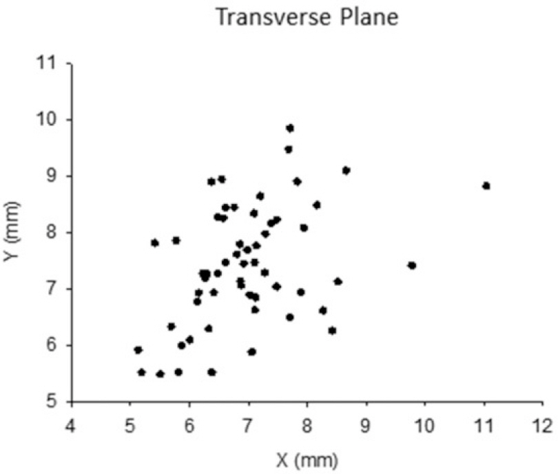
Fig. 2. Mesiodistal length (X) of incisal group in transverse plane is (6.98 ± 1.05 mm, Max = 11.05 mm), buccolingual length (Y) is (7.39 ± 1.05 mm, Max = 9.84 mm).
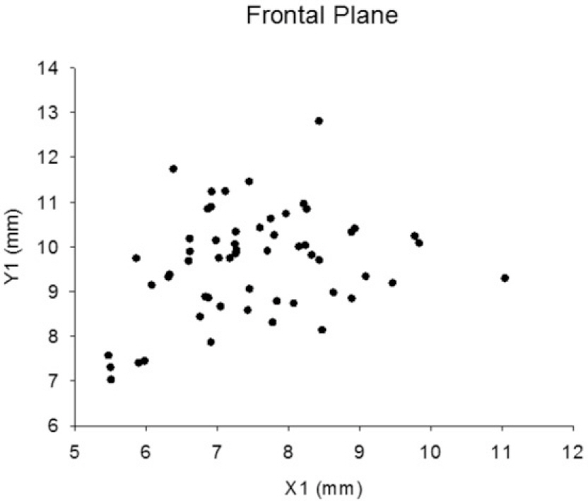
Fig. 3. Mesiodistal length (X1) of incisal group in frontal plane is (7.50 ± 1.15 mm, Max = 11.04 mm), crown height (Y) is (9.61 ± 1.17 mm, Max = 12.80 mm).
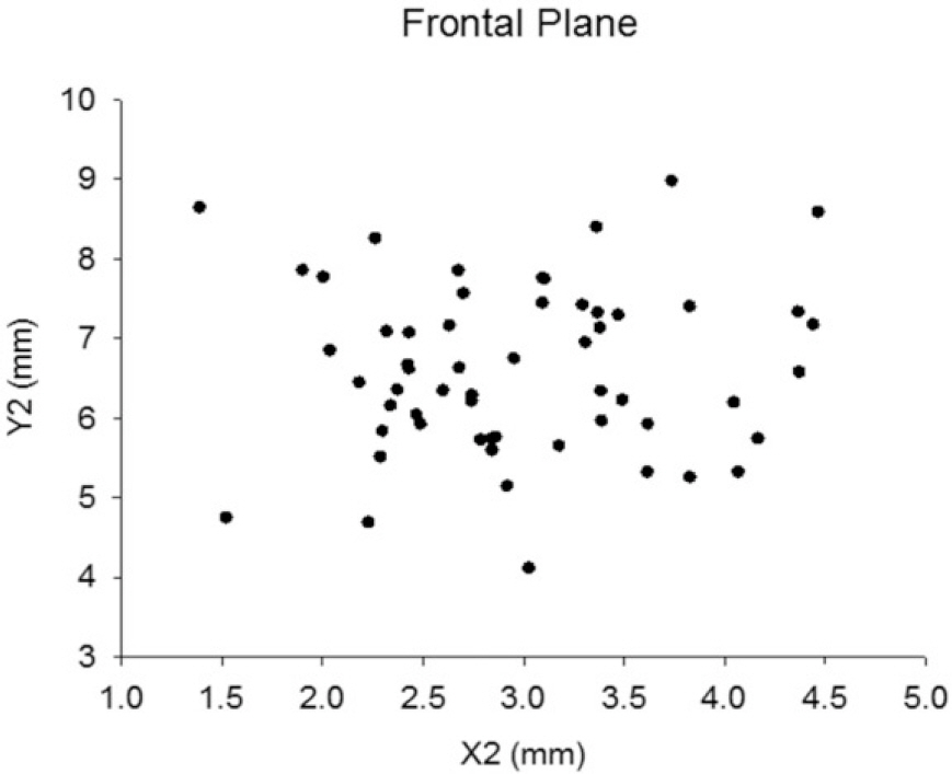
Fig. 4. Mesiodistal length (X2) of prepared incisal group in frontal plane is (2.98 ± 0.73 mm, Min = 1.39 mm), crown height (Y2) is (6.60 ± 1.05 mm, Min = 4.11 mm).
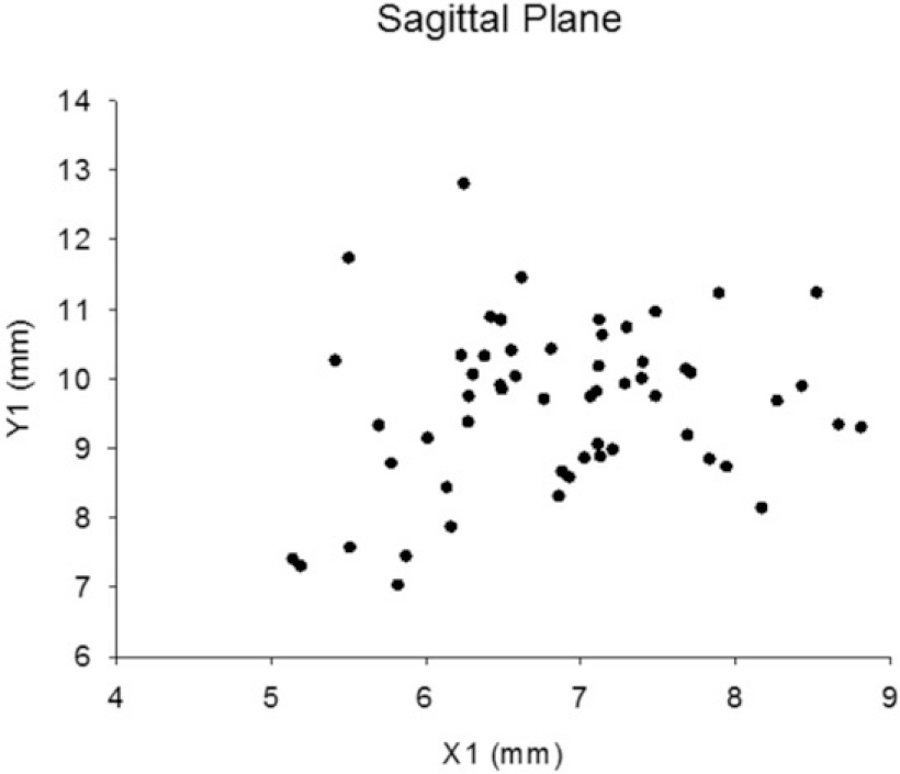
Fig. 5. Buccolingual lenth (X1) of incisal group in sagittal plane is (6.88 ± 0.88 mm, Max = 4.04 mm), crown height (Y1) is (9.61 ± 1.17 mm, Max = 9.62 mm).
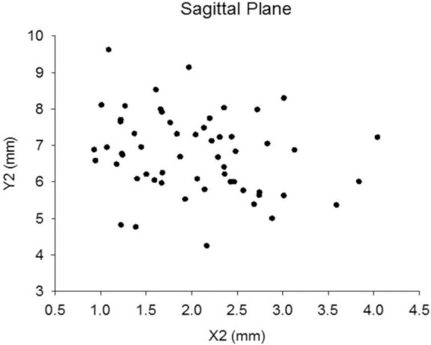
Fig. 6. Buccolingual lenth (X2) of prepared incisal group in sagittal plane is (2.04 ± 0.73 mm, Min = 0.93 mm), crown height (Y2) is (6.75 ± 1.09 mm, Min = 4.24 mm).
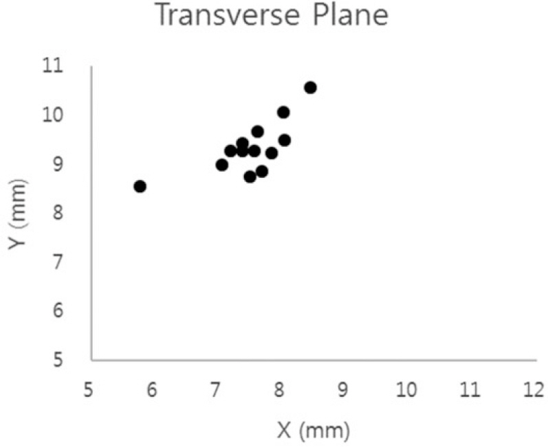
Fig. 7. Mesiodistal length (X) of premolar group in transverse plane is (8.01 ±1.47 mm, Max = 12.03 mm), buccolingual length (Y) is (9.74 ±1.18 mm, Max = 13.33 mm).
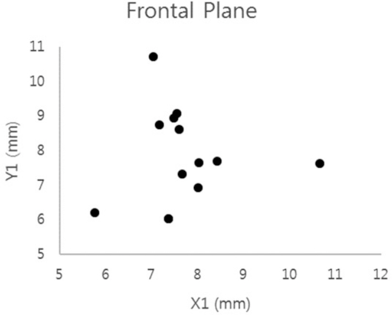
Fig. 8. Mesiodistal length (X1) of premolar group in frontal plane is (8.01 ± 1.46 mm, Max = 12.09 mm), crown height (Y) is (8.01 ± 1.48 mm, Max = 11.07 mm).
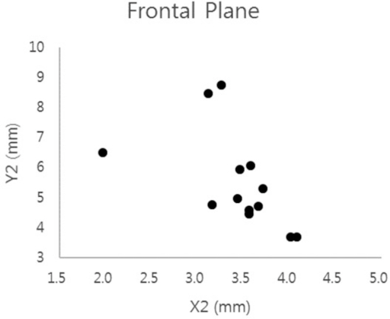
Fig. 9. Mesiodistal length (X2) of prepared premolar group in frontal plane is (4.10 ± 1.78 mm, Min = 1.97 mm), crown height (Y2) is (5.37 ± 1.49 mm, Min = 3.67 mm).
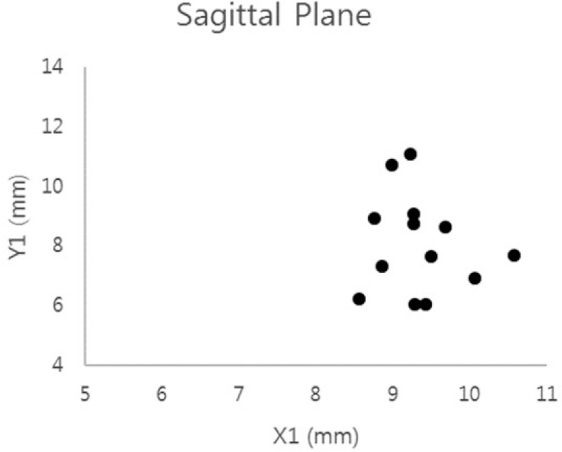
Fig. 10. Buccolingual lenth (X1) of premolar group in sagittal plane is (9.76 ± 1.22 mm, Max = 13.48 mm), crown height (Y1) is (8.01 ± 1.48 mm, Max = 11.07 mm).
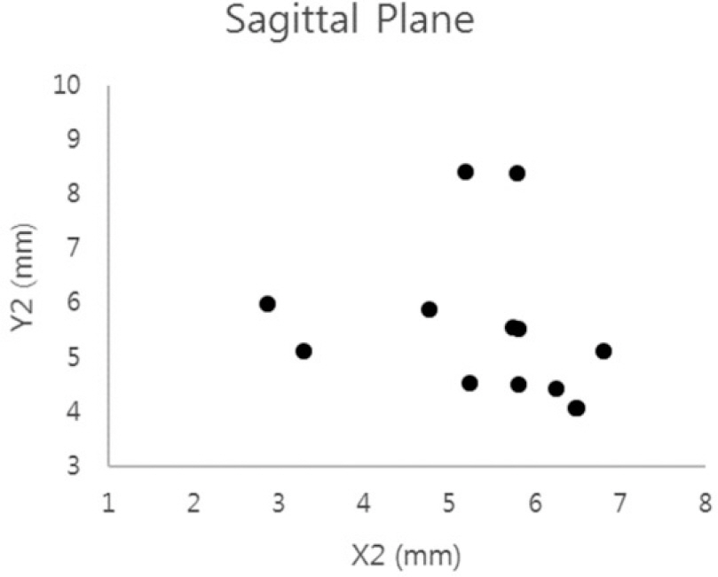
Fig. 11. Buccolingual lenth (X2) of prepared premolar group in sagittal plane is (5.86 ± 1.55 mm, Min = 2.86 mm), crown height (Y2) is (5.38 ± 1.33 mm, Min = 4.07 mm).
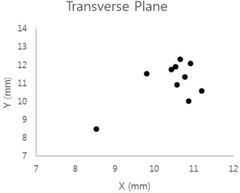
Fig. 12. Mesiodistal length (X) of molar group in transverse plane is (10.87 ± 1.04 mm, Max = 12.63 mm), buccolingual length (Y) is (11.41 ± 1.18 mm, Max = 13.33 mm).
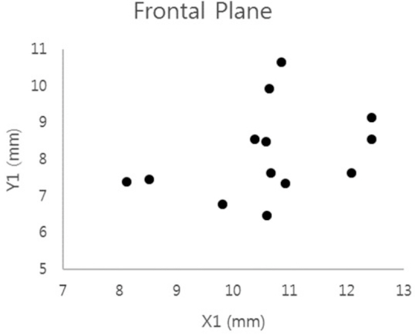
Fig. 13. Mesiodistal length (X1) of molar group in frontal plane is (10.62 ± 1.25 mm, Max = 12.44 mm), crown height (Y) is (8.15 ± 1.17 mm, Max = 10.65 mm).
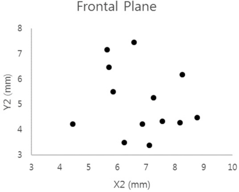
Fig. 14. Mesiodistal length (X2) of prepared molar group in frontal plane is (6.80 ± 1.18 mm, Min = 4.44 mm), crown height (Y2) is (5.11 ± 1.29 mm, Min = 3.38 mm).
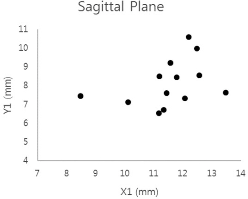
Fig. 15. Buccolingual lenth (X1) of molar group in sagittal plane is (11.53 ± 1.18 mm, Max = 13.48 mm), crown height (Y1) is (8.12 ± 1.19 mm, Max = 10.59 mm).
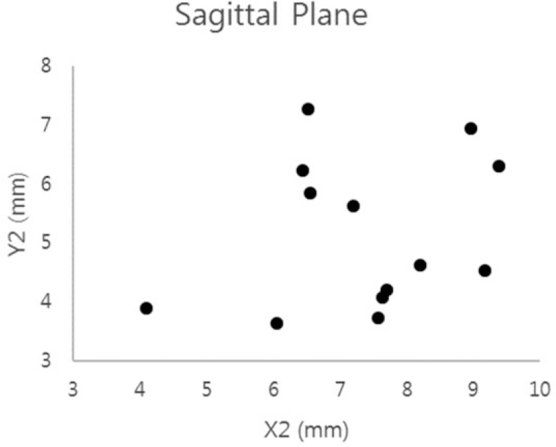
Fig. 16. Buccolingual lenth (X2) of prepared molar group in sagittal plane is (7.34 ± 1.40 mm, Min = 4.09 mm), crown height (Y2) is (5.15 ± 1.23 mm, Min = 3.63 mm).
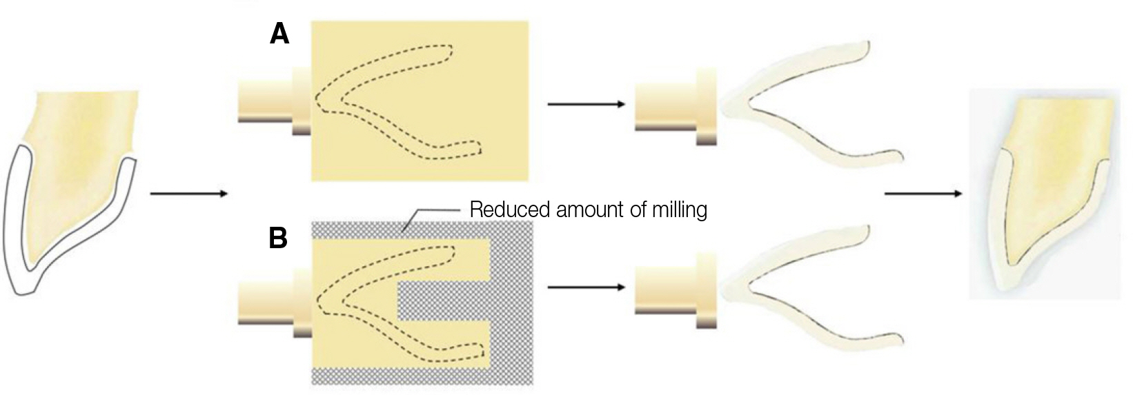
Fig. 17. Comparison of milling area between conventional zirconia block (A) and customized zirconia block (B).
Reference
-
1.Giordano RA. Dental ceramic restorative systems. Compend Contin Educ Dent. 1996. 17:779–82. 784-6 passim; quiz 794.2.Campbell SD. A comparative strength study of metal ceramic and all-ceramic esthetic materials: modulus of rupture. J Prosthet Dent. 1989. 62:476–9.
Article3.Seghi RR., Sorensen JA. Relative flexural strength of six new ceramic materials. Int J Prosthodont. 1995. 8:239–46.4.Kelly JR. Dental ceramics: current thinking and trends. Dent Clin North Am. 2004. 48(viii):513–30.5.Kang DW. Fixed prosthodontics. 1st ed.DanhanNarae Publishing: Seoul;2012. p. 330.6.Okutan M., Heydecke G., Butz F., Strub JR. Fracture load and marginal fit of shrinkage-free ZrSiO4 all-ceramic crowns after chewing simulation. J Oral Rehabil. 2006. 33:827–32.7.Hjerppe J., Vallittu PK., Fröberg K., Lassila LV. Effect of sintering time on biaxial strength of zirconium dioxide. Dent Mater. 2009. 25:166–71.
Article8.Kunii J., Hotta Y., Tamaki Y., Ozawa A., Kobayashi Y., Fujishima A., Miyazaki T., Fujiwara T. Effect of sintering on the marginal and internal fit of CAD/CAM-fabricated zirconia frameworks. Dent Mater J. 2007. 26:820–6.
Article9.Bolton WA. The clinical application of a tooth size analysis. Am J Orthod. 1962. 48:504–29.10.Kim EJ., Hwang HS. Reproducibility and accuracy of tooth size measurements obtained by the use of computer. Korean J Orthod. 1999. 29:563–73.11.Sim EJ., Hwang HS., Moon JD. A study on the error of tooth size measurements. Korean J Orthod. 1999. 29:491–502.12.Kim DS., Kim YJ., Choi JH., Han JH. A study of Korean norm about tooth size and ratio in Korean adults with normal occlusion. Korean J Orthod. 2001. 31:505–15.13.Lee SP. Dental anatomy. 1st ed.DanhanNarae Publishing: Seoul;2009. p. 46.
- Full Text Links
-
- Actions
-
Cited
- CITED
-
- Close
- Share
-
- Similar articles
-
- Colorimetric Analysis of Preformed Zirconia Anterior Crowns for Esthetic Restoration
- A full-mouth rehabilitation using zirconia all-ceramic crowns: a case report
- Comparison of marginal and internal fit of zirconia abutments with titanium abutments in internal hexagonal implants
- Fabrication of surveyed crown and repairing the artificial teeth for existing removable partial denture using digital technology: a case report
- Full-mouth rehabilitation with CAD/CAM monolithic zirconia in dentinogenesis imperfecta: a case report
