Effect of the amount of thickness reduction on color and translucency of dental monolithic zirconia ceramics
- Affiliations
-
- 1Comprehensive Treatment Center, Seoul National University Dental Hospital, Seoul, Republic of Korea.
- 2Department of Prosthodontics and Dental Research Institute, School of Dentistry, Seoul National University, Seoul, Republic of Korea. ksh1250@snu.ac.kr
- 3Department of Dentistry, Ajou University School of Medicine, Suwon, Republic of Korea.
- KMID: 2393194
- DOI: http://doi.org/10.4047/jap.2016.8.1.37
Abstract
- PURPOSE
This study investigated the effect of amount of thickness reduction on color and translucency of dental monolithic zirconia ceramics.
MATERIALS AND METHODS
One-hundred sixty-five monolithic zirconia specimens (16.3 mm x 16.3 mm x 2.0 mm) were divided into 5 groups (Group I to V) according to the number of A2-coloring liquid applications. Each group was then divided into 11 subgroups by reducing the thickness up to 1.0 mm in 0.1-mm increments (Subgroup 0 to 10, n=3). Colors and spectral distributions were measured according to CIELAB on a reflection spectrophotometer. All measurements were performed on five different areas of each specimen. Color difference (DeltaE*(ab)) and translucency parameter (TP) were calculated. Data were analyzed using one-way ANOVA and multiple comparison Scheffe test (alpha=.05).
RESULTS
There were significant differences in CIE L* between Subgroup 0 and other subgroups in all groups. CIE a* increased (0.52
CONCLUSION
Increasing thickness reduction reduces lightness and increases a reddish, bluish appearance, and translucency of monolithic zirconia ceramics.
Keyword
MeSH Terms
Figure
-
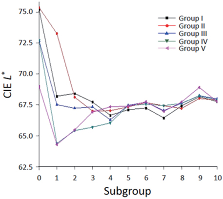
Fig. 1 Means of CIE L* values of each group against a black background as a function of the amount of thickness reduction.
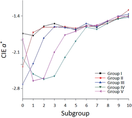
Fig. 2 Means of CIE a* values of each group against a black background as a function of the amount of thickness reduction.
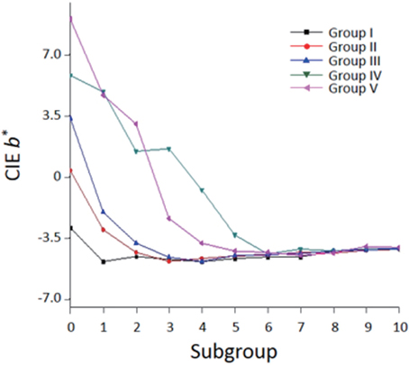
Fig. 3 Means of CIE b* values of each group against a black background as a function of the amount of thickness reduction.
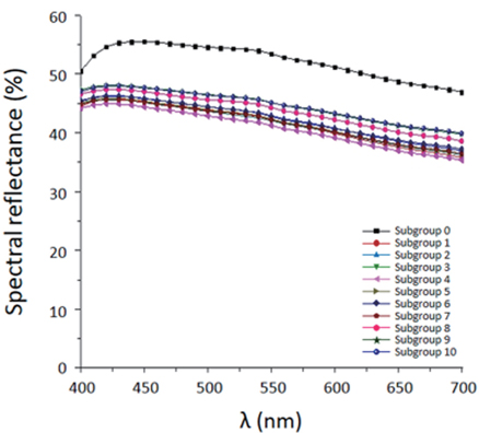
Fig. 4 Average spectral reflectance curve of each subgroup in Group I.
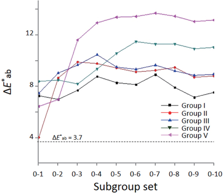
Fig. 5 Means of ΔE*ab units between Subgroup 0 and other subgroups for each group.

Fig. 6 Average spectral transmittance curve of each subgroup in Group I.
Cited by 2 articles
-
Comparative color and surface parameters of current esthetic restorative CAD/CAM materials
Ferhan Egilmez, Gulfem Ergun, Isil Cekic-Nagas, Pekka Kalevi Vallittu, Lippo Veli Juhana Lassila
J Adv Prosthodont. 2018;10(1):32-42. doi: 10.4047/jap.2018.10.1.32.Choice of resin cement shades for a high-translucency zirconia product to mask dark, discolored or metal substrates
Shiqi Dai, Chen Chen, Mo Tang, Ying Chen, Lu Yang, Feng He, Bingzhuo Chen, Haifeng Xie
J Adv Prosthodont. 2019;11(5):286-296. doi: 10.4047/jap.2019.11.5.286.
Reference
-
1. McLean JW, Odont D. Evolution of dental ceramics in the twentieth century. J Prosthet Dent. 2001; 85:61–66.2. Douglas RD, Przybylska M. Predicting porcelain thickness required for dental shade matches. J Prosthet Dent. 1999; 82:143–149.3. Isgrò G, Pallav P, van der Zel JM, Feilzer AJ. The influence of the veneering porcelain and different surface treatments on the biaxial flexural strength of a heat-pressed ceramic. J Prosthet Dent. 2003; 90:465–473.4. Tinschert J, Natt G, Mautsch W, Augthun M, Spiekermann H. Fracture resistance of lithium disilicate-, alumina-, and zirconia-based three-unit fixed partial dentures: A laboratory study. Int J Prosthodont. 2001; 14:231–238.5. Al-Amleh B, Lyons K, Swain M. Clinical trials in zirconia: a systematic review. J Oral Rehabil. 2010; 37:641–652.6. Ha SR, Kim SH, Han JS, Yoo SH, Jeong SC, Lee JB, Yeo IS. The influence of various core designs on stress distribution in the veneered zirconia crown: a finite element study. J Adv Prosthodont. 2013; 5:187–197.7. Kim HK, Kim SH, Lee JB, Han JS, Yeo IS. Effect of polishing and glazing on the color and spectral distribution of monolithic zirconia. J Adv Prosthodont. 2013; 5:296–304.8. de Azevedo Cubas GB, Camacho GB, Demarco FF, Pereira-Cenci T. The effect of luting agents and ceramic thickness on the color variation of different ceramics against a chromatic background. Eur J Dent. 2011; 5:245–252.9. Vichi A, Ferrari M, Davidson CL. Influence of ceramic and cement thickness on the masking of various types of opaque posts. J Prosthet Dent. 2000; 83:412–417.10. Shokry TE, Shen CS, Elhosary MM, Elkhodary AM. Effect of core and veneer thicknesses on the color parameters of two all-ceramic systems. J Prosthet Dent. 2006; 95:124–129.11. Dozic A, Kleverlaan CJ, Meegdes M, van der Zel J, Feilzer AJ. The influence of porcelain layer thickness on the final shade of ceramic restorations. J Prosthet Dent. 2003; 90:563–570.12. Terada Y, Maeyama S, Hirayasu R. The influence of different thicknesses of dentin porcelain on the color reflected from thin opaque porcelain fused to metal. Int J Prosthodont. 1989; 2:352–356.13. Ozturk O, Uludag B, Usumez A, Sahin V, Celik G. The effect of ceramic thickness and number of firings on the color of two all-ceramic systems. J Prosthet Dent. 2008; 100:99–106.14. Antonson SA, Anusavice KJ. Contrast ratio of veneering and core ceramics as a function of thickness. Int J Prosthodont. 2001; 14:316–320.15. Lund PS, Aquilino SA, Dixon DL. Evaluation of the color and appearance of a new textured opaque porcelain. Int J Prosthodont. 1991; 4:548–554.16. Kim HK, Kim SH. Effect of the number of coloring liquid applications on the optical properties of monolithic zirconia. Dent Mater. 2014; 30:e229–e237.17. Commission Internationale de l'Eclairage (CIE). Colorimetry, CIE 015. 3rd ed. Vienna: CIE Central Bureau;2004.18. Johnston WM, Ma T, Kienle BH. Translucency parameter of colorants for maxillofacial prostheses. Int J Prosthodont. 1995; 8:79–86.19. Johnston WM, Kao EC. Assessment of appearance match by visual observation and clinical colorimetry. J Dent Res. 1989; 68(5):819–822.20. Jorgenson MW, Goodkind RJ. Spectrophotometric study of five porcelain shades relative to the dimensions of color, porcelain thickness, and repeated firings. J Prosthet Dent. 1979; 42:96–105.21. Jacobs SH, Goodacre CJ, Moore BK, Dykema RW. Effect of porcelain thickness and type of metal-ceramic alloy on color. J Prosthet Dent. 1987; 57:138–145.22. Judd DB, Wyszecki G. Color in business, science and industry. 3rd ed. New York: John Wiley & Sons;1975. p. 397–417.23. Douglas RD, Brewer JD. Acceptability of shade differences in metal ceramic crowns. J Prosthet Dent. 1998; 79:254–260.24. Heffernan MJ, Aquilino SA, Diaz-Arnold AM, Haselton DR, Stanford CM, Vargas MA. Relative translucency of six all-ceramic systems. Part I: Core materials. J Prosthet Dent. 2002; 88:4–9.25. Heffernan MJ, Aquilino SA, Diaz-Arnold AM, Haselton DR, Stanford CM, Vargas MA. Relative translucency of six all-ceramic systems. Part II: Core and veneer materials. J Prosthet Dent. 2002; 88:10–15.26. O'Keefe KL, Pease PL, Herrin HK. Variables affecting the spectral transmittance of light through porcelain veneer samples. J Prosthet Dent. 1991; 66:434–438.27. Nassau K. The physics and chemistry of color. 2nd ed. New York: John Wiley & Sons;2001. p. 231–236. p. 39028. Kingery WD, Bowen HK, Uhlmann DR. Introduction to ceramic. 2nd ed. New York: John Wiley & Sons;1976. p. 668.
- Full Text Links
-
- Actions
-
Cited
- CITED
-
- Close
- Share
-
- Similar articles
-
- Comparison of the optical properties of pre-colored dental monolithic zirconia ceramics sintered in a conventional furnace versus a microwave oven
- Esthetic anterior restoration using 3M Lavaâ„¢ Esthetic monolithic zirconia
- Influences of luting cement shade on the color of various translucent monolithic zirconia and lithium disilicate ceramics for veneer restorations
- Comparison of the translucency of shaded zirconia all-ceramic systems
- Evaluation of translucency of monolithic zirconia and framework zirconia materials
