Investig Magn Reson Imaging.
2017 Sep;21(3):195-198. 10.13104/imri.2017.21.3.195.
Magnetic Resonance Imaging of Idiopathic Herniation of the Lingual Gyrus: a Case Report
- Affiliations
-
- 1Department of Radiology, Seoul Medical Center, Seoul, Korea. jnoon276@gmail.com
- KMID: 2392693
- DOI: http://doi.org/10.13104/imri.2017.21.3.195
Abstract
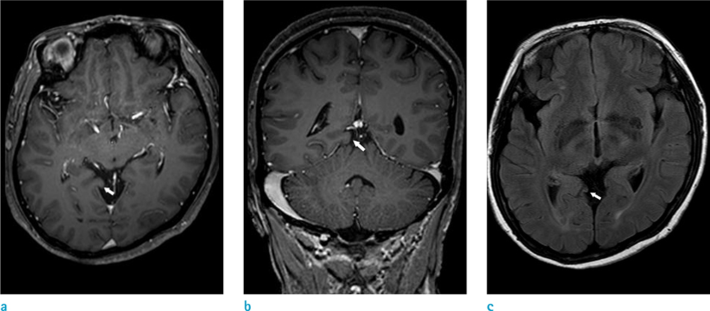
- Idiopathic brain herniation is a rare condition. We believe that this is the first reported case of idiopathic herniation of the lingual gyrus. The case involves a 57-year-old woman presenting with frontal headache without overt visual symptoms. Magnetic resonance imaging (MRI) revealed an idiopathic herniation of the lingual gyrus of the occipital lobe extending into the quadrigeminal cistern. No other adjacent intracranial abnormalities were observed. Although some conditions may be considered in the differential diagnosis, accurate diagnosis of idiopathic brain herniation in medical practice can prevent unnecessary additional imaging procedures and invasive open biopsy in patients with typical imaging findings.
MeSH Terms
Figure
Reference
-
1. Maldjian C, Adam R. Prevalence of idiopathic cuneate gyrus herniation based on emergency room CT examinations. Emerg Radiol. 2014; 21:387–389.2. Duarte MP, Maldjian TC, Tenner M, Adam R. Magnetic resonance imaging of idiopathic herniation of the cuneus gyrus. J Neuroimaging. 2007; 17:353–354.3. Koc G, Doganay S, Bayram AK, et al. Idiopathic brain herniation. A report of two paediatric cases. Neuroradiol J. 2014; 27:586–589.4. Horowitz M, Kassam A, Levy E, Lunsford LD. Misinterpretation of parahippocampal herniation for a posterior fossa tumor: imaging and intraoperative findings. J Neuroimaging. 2002; 12:78–79.5. Yavarian Y, Bayat M, Brondum Frokjaer J. Herniation of uncus and parahippocampal gyrus: an accidental finding on magnetic resonance imaging of cerebrum. Acta Radiol Short Rep. 2015; 4:2047981614560077.6. Blumenfeld H. Neuroanatomy through clinical cases. Disorders of higher-order visual processing. Sunderland, MA: Sinauer Associates, Inc.;2010. p. 914–917.7. Larsen WJ. Human embryology. New York; Edinburgh: Churchill Livingstone;1997. p. 435–437.8. O'Rahilly R, Muller F. The meninges in human development. J Neuropathol Exp Neurol. 1986; 45:588–608.
- Full Text Links
- Actions
-
Cited
- CITED
-
- Close
- Share
- Similar articles
-
- Idiopathic Spinal Cord Herniation Presented as Brown-Sequard Syndrome: A Case Report and Surgical Outcome
- Spinal Nerve Root Swelling Mimicking Intervertebral Disc Herniation in Magnetic Resonance Imaging: A Case Report
- A Lumbar Disc Herniation Misdiagnosed as A Neurofibromatosis Type I: A Case Report
- The Relationship between the Lower Lumbar Disc Herniation and the Morphology of the Iliolumbar Ligaments Using Magnetic Resonance Imaging
- Thiemann's Disease: a Case Report


