Giant Arachnoid Granulations in Headache Mimicking Migraine with Aura
- Affiliations
-
- 1Department of Neurology, Dongguk University Ilsan Hospital, Goyang, Korea. noizegun@gmail.com
- 2Department of Neuroradiology, Dongguk University Ilsan Hospital, Goyang, Korea.
- KMID: 2392692
- DOI: http://doi.org/10.13104/imri.2017.21.3.192
Abstract
- Giant arachnoid granulations have been reported to be associated with headaches, which can be acute or chronic in presentation. In some cases, idiopathic intracranial hypertension, previously called pseudotumor cerebri, may occur. The pathophysiology of these enlarged structures seen as filling defects on imaging is not clearly defined, although they are presumed to cause symptoms such as headache via pressure resulting from secondary venous sinus obstruction. We present a unique presentation of secondary headache in a 39-year-old man with no prior history of headaches found to have giant arachnoid granulations, presenting as migraine with aura.
Keyword
MeSH Terms
Figure
-
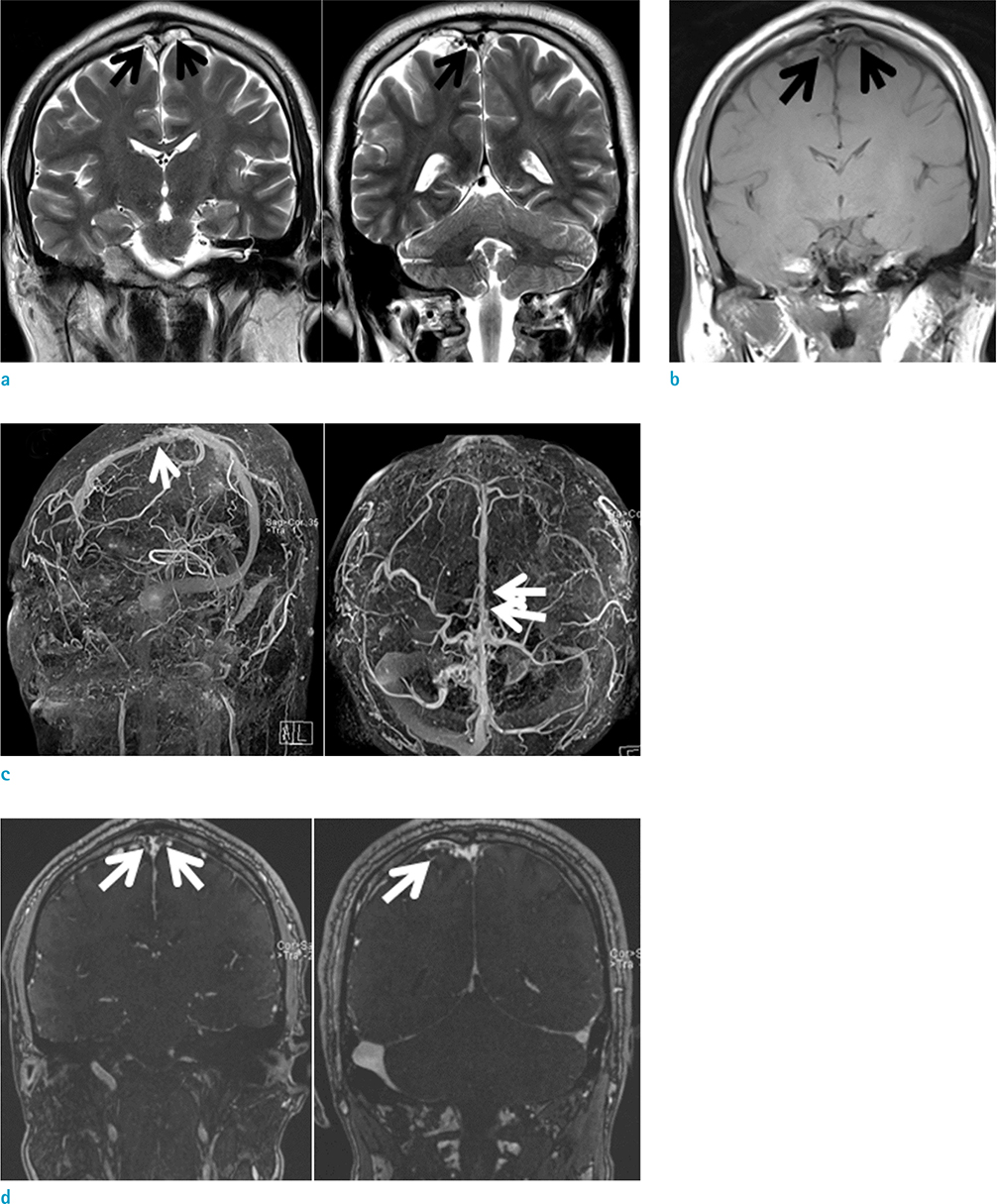
Fig. 1 (a) Coronal T2-weighted image. Multifocal amorphous hyperintense lesions compressing the superior sagittal sinus are seen (black arrows). Tubular hyperintense lesion (short black arrow) in the right superficial cortical vein is also seen. (b) Coronal T1-weighted image. Hypointense lesions (black arrows) are noted. (c) Contrast-enhanced MR venography. Irregular narrowing or the lumen and multifocal filling defects in the superior sagittal sinuses (long white arrows) as well as the right superficial cortical vein (short white arrow) are noted. (d) Coronal source imaging of MR venography. There are poorly opacified lesions (white arrows) compressing the superior sagittal sinus. Filling defects (white arrow) in the right superficial cortical vein are seen.
Reference
-
1. Arjona A, Delgado F, Fernandez-Romero E. Intracranial hypertension secondary to giant arachnoid granulations. J Neurol Neurosurg Psychiatry. 2003; 74:418.2. Kiroglu Y, Yaqci B, Cirak B, Karabulut N. Giant arachnoid granulation in a patient with benign intracranial hypertension. Eur Radiol. 2008; 18:2329–2332.3. Zheng H, Zhou M, Zhao B, Zhou D, He L. Pseudotumor cerebri syndrome and giant arachnoid granulation: treatment with venous sinus stenting. J Vasc Interv Radiol. 2010; 21:927–929.4. Peters SA, Frombach E, Heyer CM. Giant arachnoid granulation: differential diagnosis of acute headache. Australas Radiol. 2007; 51 Spec No.:B18–B20.5. Choi HJ, Cho CW, Kim YS, Cha JH. Giant arachnoid granulation misdiagnosed as transverse sinus thrombosis. J Korean Neurosurg Soc. 2008; 43:48–50.6. Kan P, Stevens EA, Couldwell WT. Incidental giant arachnoid granulation. AJNR Am J Neuroradiol. 2006; 27:1491–1492.
- Full Text Links
-
- Actions
-
Cited
- CITED
-
- Close
- Share
-
- Similar articles
-
- Clinical Characteristics of Migraine with Aura in Korean: a Clinic Based Study
- A Case of Successful Treatment During Migraine Aura Using Isometheptene Compound
- Persistent Aura Without Infarction
- Typical Aura without Headache Presenting Intermittent Transient Visual Symptom
- The Diagnosis and Most-Updated Therapy of Migraine
