A Rare Case of Intramural Müllerian Adenosarcoma Arising from Adenomyosis of the Uterus
- Affiliations
-
- 1Department of Pathology, Catholic University of Daegu School of Medicine, Daegu, Korea. pathosjlee@cu.ac.kr
- KMID: 2392586
- DOI: http://doi.org/10.4132/jptm.2017.06.11
Abstract
- Müllerian adenosarcomas usually arise as polypoid masses in the endometrium of post-menopausal women. Occasionally, these tumors arise in the cervix, vagina, broad and round ligaments, ovaries and rarely in extragenital sites; these cases are generally associated with endometriosis. We experienced a rare case of extraendometrial, intramural adenosarcoma arising in a patient with adenomyosis. A 40-year-old woman presented with sudden-onset suprapubic pain. The imaging findings suggested leiomyoma with cystic degeneration in the uterine fundus. An ill-defined ovoid tumor with hemorrhagic degeneration, measuring 7.5 cm in diameter, was detected. The microscopic findings showed glandular cells without atypia and a sarcomatous component with pleomorphism and high mitotic rates. There was no evidence of endometrial origin. To recognize that adenosarcoma can, although rarely, arise from adenomyosis is important to avoid overstaging and inappropriate treatment.
Keyword
MeSH Terms
Figure
-
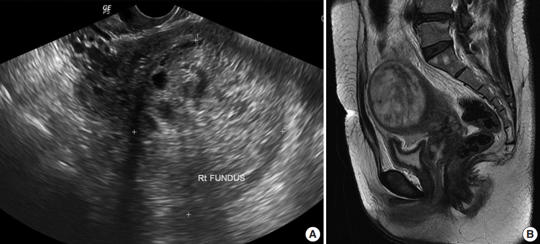
Fig. 1. Radiologic findings. (A) Pelvic ultrasonography, showing an enlarged uterus with a 7-cm solid and cystic mass. (B) T2-weighted magnetic resonance imaging, showing an enlarged uterus with a mass, measuring 71×59×72 mm, arising from the uterine fundus.
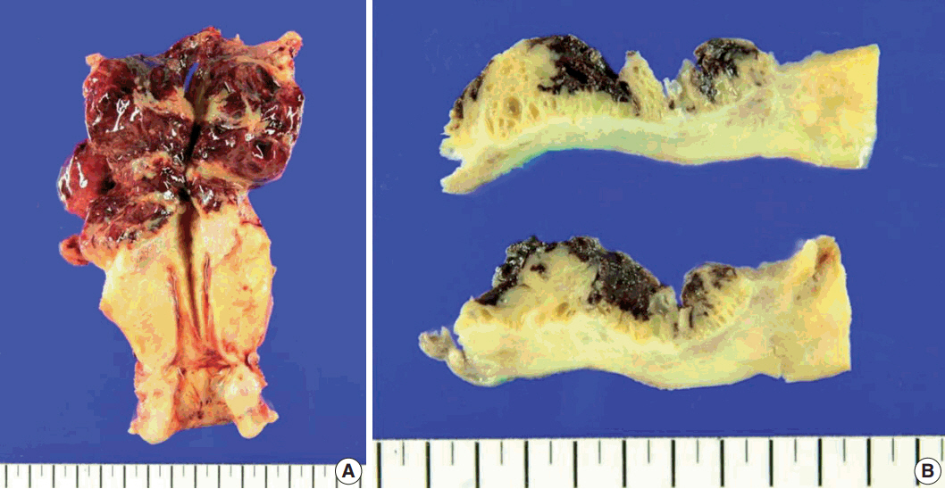
Fig. 2. Macroscopic findings of the hysterectomy specimen. (A) View showing an ill-defined ovoid tumor, 7.5 cm in diameter, together with hemorrhagic degeneration in the uterine fundus. (B) View showing that the cut surface of the lesion was tan-brown in color, multicystic, and solid.

Fig. 3. Histologic and immunohistochemical findings. (A, B) Microscopic examination showing a biphasic tumor composed of both dilated glandular elements and abundant, hypercellular stromal elements. (C) The tumor shows expansile growth within the myometrium, with extensive myometrial invasion and focal infiltrates into the subserosa with expansile margins. (D) Proliferation of hypercellular spindle cells growing in a fascicular pattern, around benign endometrial glands. (E) The stromal cells show mild and focal moderate cytological atypia with occasional mitotic figures (arrows). (F–J) Immunohistochemical analysis showing that the glandular and stromal cells in tumor tissue are positive for estrogen receptor (F) and focally positive for p53 (G); that the stromal cells are positive for CD10 (H) and smooth muscle actin (I); and that the Ki-67 proliferation index is higher in the stromal component than in the epithelium (J).
Reference
-
1. Pinto A, Howitt B. Uterine adenosarcoma. Arch Pathol Lab Med. 2016; 140:286–90.
Article2. Elshafie M, Rahimi S, Ganesan R, Hirschowitz L. Müllerian adenosarcoma arising in a subserosal adenomyoma. Int J Surg Pathol. 2013; 21:186–9.
Article3. McCluggage WG. Müllerian adenosarcoma of the female genital tract. Adv Anat Pathol. 2010; 17:122–9.4. Raffaelli R, Piazzola E, Zanconato G, Fedele L. A rare case of extrauterine adenosarcoma arising in endometriosis of the rectovaginal septum. Fertil Steril. 2004; 81:1142–4.
Article5. Gollard R, Kosty M, Bordin G, Wax A, Lacey C. Two unusual presentations of mullerian adenosarcoma: case reports, literature review, and treatment considerations. Gynecol Oncol. 1995; 59:412–22.6. Verschraegen CF, Vasuratna A, Edwards C, et al. Clinicopathologic analysis of Müllerian adenosarcoma: the M.D. Anderson Cancer Center experience. Oncol Rep. 1998; 5:939–44.7. Liu L, Davidson S, Singh M. Müllerian adenosarcoma of vagina arising in persistent endometriosis: report of a case and review of the literature. Gynecol Oncol. 2003; 90:486–90.
Article8. Clarke BA, Mulligan AM, Irving JA, McCluggage WG, Oliva E. Müllerian adenosarcomas with unusual growth patterns: staging issues. Int J Gynecol Pathol. 2011; 30:340–7.9. Prat J. FIGO staging for uterine sarcomas. Int J Gynaecol Obstet. 2009; 104:177–8.
Article10. Jha P, Ansari C, Coakley FV, et al. Case report: Imaging of Müllerian adenosarcoma arising in adenomyosis. Clin Radiol. 2009; 64:645–8.11. Major FJ, Blessing JA, Silverberg SG, et al. Prognostic factors in early-stage uterine sarcoma: a Gynecologic Oncology Group study. Cancer. 1993; 71(4 Suppl):1702–9.
Article12. Abeler VM, Royne O, Thoresen S, Danielsen HE, Nesland JM, Kristensen GB. Uterine sarcomas in Norway: a histopathological and prognostic survey of a total population from 1970 to 2000 including 419 patients. Histopathology. 2009; 54:355–64.
Article13. Early HM, McGahan JP, Naderi S, Lamba R, Fananapazir G. Mullerian adenosarcoma: a malignant progression of adenomyosis? Pictorial review with multimodality imaging. J Ultrasound Med. 2015; 34:2109–13.14. Bocklage T, Lee KR, Belinson JL. Uterine Müllerian adenosarcoma following adenomyoma in a woman on tamoxifen therapy. Gynecol Oncol. 1992; 44:104–9.15. Oda Y, Nakanishi I, Tateiwa T. Intramural Müllerian adenosarcoma of the uterus with adenomyosis. Arch Pathol Lab Med. 1984; 108:798–801.16. Clement PB, Scully RE. Müllerian adenosarcoma of the uterus: a clinicopathologic analysis of 100 cases with a review of the literature. Hum Pathol. 1990; 21:363–81.17. Howitt BE, Sholl LM, Dal Cin P, et al. Targeted genomic analysis of Müllerian adenosarcoma. J Pathol. 2015; 235:37–49.
Article18. Chen Z, Hong B, Drozd-Borysiuk E, Coffin C, Albritton K. Molecular cytogenetic characterization of a case of Müllerian adenosarcoma. Cancer Genet Cytogenet. 2004; 148:129–32.
Article19. Gilks CB, Clement PB, Hart WR, Young RH. Uterine adenomyomas excluding atypical polypoid adenomyomas and adenomyomas of endocervical type: a clinicopathologic study of 30 cases of an underemphasized lesion that may cause diagnostic problems with brief consideration of adenomyomas of other female genital tract sites. Int J Gynecol Pathol. 2000; 19:195–205.
Article20. Gallardo A, Prat J. Müllerian adenosarcoma: a clinicopathologic and immunohistochemical study of 55 cases challenging the existence of adenofibroma. Am J Surg Pathol. 2009; 33:278–88.
- Full Text Links
-
- Actions
-
Cited
- CITED
-
- Close
- Share
-
- Similar articles
-
- A Case of Mllerian adenosarcoma of vaginal stump after total abdominal hysterectomy
- Mullerian Adenosarcoma Arising From Rectal Endometriosis
- MR Findings of Extrauterine Mullerian Adenosarcoma Associated with Deep Pelvic Endometriosis
- Adenocarcinoma Arising in Adenomyosis
- A Case of M llerian Adenosarcoma of the Uterus with Sarcomatous Overgrowth
