A Rare Case of Aggressive Melanotic Schwannoma Occurred in Spinal Nerve of a 59-Year-Old Male
- Affiliations
-
- 1Department of Pathology, Gangnam Severance Hospital, Yonsei University College of Medicine, Seoul, Korea.
- 2Department of Neurosurgery, Gangnam Severance Hospital, Yonsei University College of Medicine, Seoul, Korea.
- 3Department of Radiology, Gangnam Severance Hospital, Yonsei University College of Medicine, Seoul, Korea.
- 4Department of Pathology, Severance Hospital, Yonsei University College of Medicine, Seoul, Korea. paxco@yuhs.ac
- KMID: 2392570
- DOI: http://doi.org/10.4132/jptm.2017.01.04
Abstract
- Melanotic schwannoma (MS) is a rare variant of nerve sheath neoplasm that shows ultrastructural and immunophenotypical features of Schwann cells but also has cytoplasmic melanosomes and is reactive for melanocytic markers as well. Unlike conventional schwannoma, which is totally benign, MS has an unpredictable prognosis and is thought to have low-malignant potential. Herein, we present a rare case of recurrent MS in lumbar spine of a 59-year-old male.
Keyword
MeSH Terms
Figure
-
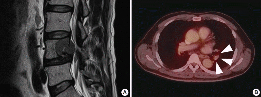
Fig. 1. Radiologic findings of spinal melanotic schwannoma. (A) Magnetic resonance imaging of lumbar spine reveals a destructive mass of the vertebral body. (B) Metastatic pulmonary nodule in left upper lobe with increased fluorodeoxyglucose uptake on positron emission tomography–computed tomography (arrowheads).
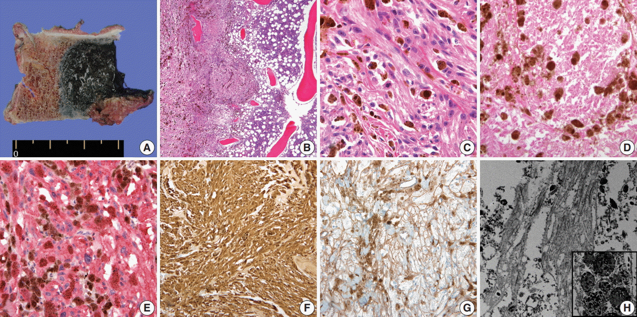
Fig. 2. Gross, microscopic and ultrastructural findings of melanotic schwannoma. (A) A heavily pigmented black round mass of vertebral body has infiltrative margin. (B) Tumor cells permeate the bone marrow space of vertebra (left). Note the right sided normal bone marrow that shows retained trabecular bone and marrow space containing hematopoietic cells. (C) Epithelioid tumor cells have discernible cytoplasmic membrane, pleomorphic nuclei, and cytoplasmic melanin pigments. Note the mitosis (center) and prominent nucleolus. (D) Foci of tumor necrosis are seen. Human melanoma black 45 (E) and S-100 protein (F) are diffusely and strongly positive in tumor cells. (G) Collagen type IV staining reveals pericellular membranous staining of tumor cells, implying the presence of basal lamina. (H) On electron microscopy, abundant basal lamina of tumor cell is evident with cytoplasmic melanosomes (×12,000) (inset, ×5,000).
Reference
-
1. Carney JA. Carney complex: the complex of myxomas, spotty pigmentation, endocrine overactivity, and schwannomas. Semin Dermatol. 1995; 14:90–8.
Article2. Louis DN, Ohgaki H, Wiestler OD, Cavenee WK. WHO classification of tumours of the central nervous system. Lyon: IARC Press;2007.3. Yim H, Go JH, Ahn CS, Hong SW, Jung WH. Pigmented (melanotic) schwannoma of the cervical spinal canal: a case report. Korean J Pathol. 1995; 29:256–62.4. Yi S, Chin DK, Jin BH, Cho YE, Kim YS. Melanotic schwannoma in cervical spine: a case report. J Korean Neurosurg Soc. 2001; 30:916–20.5. You SH, Suh YL, Kim JH. Melanotic acoustic schwannoma. J Korean Neurosurg Soc. 2002; 31:485–7.6. Carney JA. Psammomatous melanotic schwannoma: a distinctive, heritable tumor with special associations, including cardiac myxoma and the Cushing syndrome. Am J Surg Pathol. 1990; 14:206–22.7. Vallat-Decouvelaere AV, Wassef M, Lot G, et al. Spinal melanotic schwannoma: a tumour with poor prognosis. Histopathology. 1999; 35:558–66.
Article8. Torres-Mora J, Dry S, Li X, Binder S, Amin M, Folpe AL. Malignant melanotic schwannian tumor: a clinicopathologic, immunohistochemical, and gene expression profiling study of 40 cases, with a proposal for the reclassification of “melanotic schwannoma”. Am J Surg Pathol. 2014; 38:94–105.9. Kusters-Vandevelde HV, van Engen-van Grunsven IA, Kusters B, et al. Improved discrimination of melanotic schwannoma from melanocytic lesions by combined morphological and GNAQ mutational analysis. Acta Neuropathol. 2010; 120:755–64.
Article10. Santaguida C, Sabbagh AJ, Guiot MC, Del Maestro RF. Aggressive intramedullary melanotic schwannoma: case report. Neurosurgery. 2004; 55:1430.
Article11. Tawk RG, Tan D, Mechtler L, Fenstermaker RA. Melanotic schwannoma with drop metastases to the caudal spine and high expression of CD117 (c-kit). J Neurooncol. 2005; 71:151–6.
Article12. Shields LB, Glassman SD, Raque GH, Shields CB. Malignant psammomatous melanotic schwannoma of the spine: a component of Carney complex. Surg Neurol Int. 2011; 2:136.
Article13. Faria MH, Doria-Netto RH, Osugue GJ, Queiroz Lde S, Chaddad-Neto FE. Melanotic schwannoma of the cervical spine progressing with pulmonary metastasis: case report. Neurol Med Chir (Tokyo). 2013; 53:712–6.
Article14. Khoo M, Pressney I, Hargunani R, Tirabosco R. Melanotic schwannoma: an 11-year case series. Skeletal Radiol. 2016; 45:29–34.
Article15. Rowlands D, Edwards C, Collins F. Malignant melanotic schwannoma of the bronchus. J Clin Pathol. 1987; 40:1449–55.
Article16. Killeen RM, Davy CL, Bauserman SC. Melanocytic schwannoma. Cancer. 1988; 62:174–83.
Article
- Full Text Links
-
- Actions
-
Cited
- CITED
-
- Close
- Share
-
- Similar articles
-
- Melanotic Schwannoma in Cervical Spine: A Case Report
- Melanotic Acoustic Schwannoma
- Pigmented(melanotic) Schwannoma of the Cervical Spinal Canal: A case report
- A Case of Schwannoma of the Chorda Tympani Nerve
- Surgical treatment of multiple plexiform schwannomas arising from the superficial radial nerve: a case report
