Supratentorial Anaplastic Ependymoma Mimicking an Extra-Axial Tumor: A Case Report
- Affiliations
-
- 1Department of Radiology, Jeju National University Hospital, Jeju National University School of Medicine, Jeju, Korea. hoklee33@gmail.com
- 2Department of Pathology, Jeju National University Hospital, Jeju National University School of Medicine, Jeju, Korea.
- 3Department of Neurosurgery, Jeju National University Hospital, Jeju National University School of Medicine, Jeju, Korea.
- KMID: 2392124
- DOI: http://doi.org/10.3348/jksr.2017.77.4.254
Abstract
- Ependymoma is a glioma which arises from the ependymal cells lining the ventricle and the central spinal canal. It commonly occurs in the brain outside the ventricle in the supratentorium, in case of a supratentorial ependymoma. We report a case of supratentorial exophytic anaplastic ependymoma mimicking an extra-axial tumor.
Figure
-
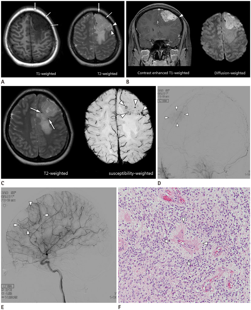
Fig. 1 A 21-year-old woman with supratentorial anaplastic ependymoma. A. Axial T1-weighted and T2-weighted images show a 5.3-cm, lobulated mass on the left frontal convexity. The mass abuts the inner surface of the skull with a broad base (arrows). The cerebrospinal fluid cleft sign is noted on the T2-weighted image (arrowheads). The mass shows heterogeneous low signal intensity on the T1-weighted image, and intermediate signal intensity on the T2-weighted image. B. The mass shows heterogenous enhancement on the gadolinium-enhanced T1-weighted fat-suppressed image, and high signal intensity on the diffusion-weighted image. Coronal contrast-enhanced T1-weighted MR image shows the dural tail sign (arrowhead). C. The interrupted surface of the left frontal lobe is considered as the epicenter of the neoplasm (arrows). The susceptibility-weighted image shows multiple dark signal intensity foci, which are suggestive of microhemorrhages (arrowheads). D. On the left external carotid angiography, the center of the tumor blush (arrowheads) is fed by the left middle meningeal artery. E. On the left internal carotid arteriography, the periphery of the tumor (arrowheads) is fed by the left anterior cerebral artery. F. On histopathology, distinct perivascular pseudorosettes (arrowheads) with the formation of an “anuclear zone” are found in some regions of the tumor, often associated with microvascular proliferation. Tumor cells have hyperchromatic, round to oval nuclei. True ependymal rosettes or ependymal canals are not detected (hematoxylin and eosin stain, × 200).
Reference
-
1. Davis MJ, Hasan F, Weinreb I, Wallace MC, Kiehl TR. Extraventricular anaplastic ependymoma with metastasis to scalp and neck. J Neurooncol. 2011; 104:599–604.2. Elsharkawy AE, Abuamona R, Bergmann M, Salem S, Gafumbegete E, Röttger E. Cortical anaplastic ependymoma with significant desmoplasia: a case report and literature review. Case Rep Oncol Med. 2013; 2013:354873.3. Singh V, Turel MK, Chacko G, Joseph V, Rajshekhar V. Supratentorial extra-axial anaplastic ependymoma mimicking a meningioma. Neurol India. 2012; 60:111–113.4. Leng X, Tan X, Zhang C, Lin H, Qiu S. Magnetic resonance imaging findings of extraventricular anaplastic ependymoma: a report of 11 cases. Oncol Lett. 2016; 12:2048–2054.5. Shuangshoti S, Rushing EJ, Mena H, Olsen C, Sandberg GD. Supratentorial extraventricular ependymal neoplasms: a clinicopathologic study of 32 patients. Cancer. 2005; 103:2598–2605.6. Vernet O, Farmer JP, Meagher-Villemure K, Montes JL. Supratentorial ectopic ependymoma. Can J Neurol Sci. 1995; 22:316–319.7. Mangalore S, Aryan S, Prasad C, Santosh V. Imaging characteristics of supratentorial ependymomas: study on a large single institutional cohort with histopathological correlation. Asian J Neurosurg. 2015; 10:276–281.8. Han MH, Park KS, Park SH, Hwang JH. Supratentorial extraventricular anaplastic ependymoma presenting with repeated intratumoral hemorrhage. Brain Tumor Res Treat. 2014; 2:81–86.9. Osborn AG. Osborn's brain: imaging, pathology and anatomy. 1st ed. Philadelphia: Lippincott Williams & Wilkins;2013. p. 585–595. .10. Sotoudeh H, Yazdi HR. A review on dural tail sign. World J Radiol. 2010; 2:188–192.
- Full Text Links
-
- Actions
-
Cited
- CITED
-
- Close
- Share
-
- Similar articles
-
- A Case of Supratentorial Intra-axial Ependymoma Showing Exophytic Growth
- Supratentorial Extraventricular Anaplastic Ependymoma Presenting with Repeated Intratumoral Hemorrhage
- A Case of Recurrent Supratentorial Extraventricular Anaplastic Ependymoma in Adult
- Gangliocytoma Mimicking Extra-axial Tumor: A Report of Two Cases
- Supratentorial Cortical Ependymoma in a 21-Month-Old Boy
