High-intensity focused ultrasound combined with hysteroscopic resection to treat retained placenta accreta
- Affiliations
-
- 1Aegis Woman Medical Clinic, Seoul, Korea.
- 2Green Cross Medical Clinic, Incheon, Korea.
- 3Department of Obstetrics and Gynecology, Incheon St. Mary's Hospital, The Catholic University of Korea College of Medicine, Incheon, Korea. tekim@catholic.ac.kr
- KMID: 2391696
- DOI: http://doi.org/10.5468/ogs.2016.59.5.421
Abstract
- We present a case of retained placenta accreta treated by high-intensity focused ultrasound (HIFU) ablation followed by hysteroscopic resection. The patient was diagnosed as submucosal myoma based on ultrasonography in local clinic. Pathologic examination of several pieces of tumor mass from the hysteroscopic procedure revealed necrotic chorionic villi with calcification. HIFU was performed using an ultrasound-guided HIFU tumor therapeutic system. The ultrasound machine had been used for real-time monitoring of the HIFU procedure. After HIFU treatment, no additional vaginal bleeding or complications were observed. A hysteroscopic resection was performed to remove ablated placental tissue 7 days later. No abnormal vaginal bleeding or discharge was seen after the procedure. The patient was stable postoperatively. We proposed HIFU and applied additional hysteroscopic resection for a safe and effective method for treating retained placenta accreta to prevent complications from the remaining placental tissue and to improve fertility options.
Keyword
MeSH Terms
Figure
-
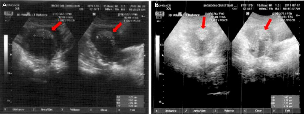
Fig. 1 Pre and post 1st hysteroscopic resection sono finding. (A) Transabdominal sonographic finding before 1st hysteroscopic resection (coronal and sagittal view): 3.2×2.0×1.5-cm hyperechogenic shadow in the endometrial cavity was shown (arrow). (B) Transabdominal sonographic finding after 1st hysteroscopic resection (coronal and sagittal view): remained hyperechogenic shadow was shown (arrow).
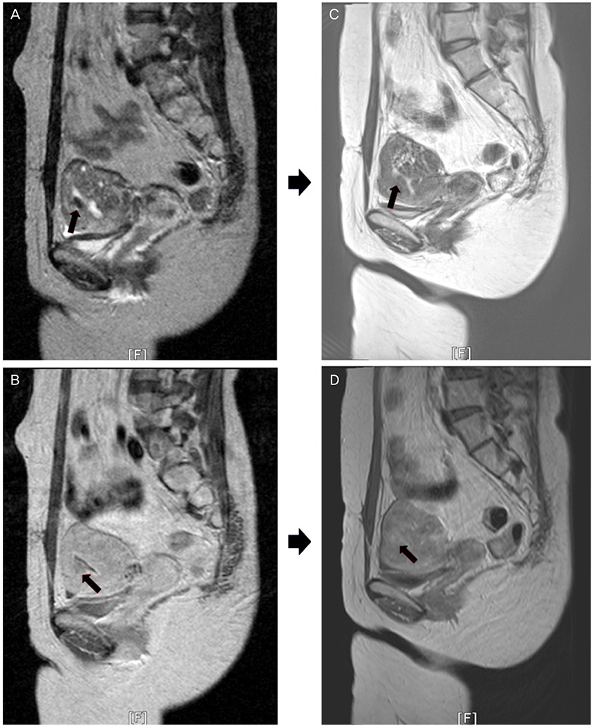
Fig. 2 Pre and post high-intensity focused ultrasound (HIFU) combined with 2nd hysteroscopic resection magnetic resonance imaging finding (MRI). (A) T2W1 and (B) contrast enhanced image was MRI finding before HIFU ablation. The retained placenta accreta was shown as low signal intensity on (A) and low enhanced image on (B). (C) T2W1 and (D) contrast enhanced image was MRI finding 3 months later after HIFU combined with 2nd hysteroscopic resection. The lesion was not seen.
Reference
-
1. Committee on Obstetric Practice. Committee opinion no. 529: placenta accreta. Obstet Gynecol. 2012; 120:207–211.2. Tanaka YO, Shigemitsu S, Ichikawa Y, Sohda S, Yoshikawa H, Itai Y. Postpartum MR diagnosis of retained placenta accreta. Eur Radiol. 2004; 14:945–952.3. Verspyck E, Resch B, Sergent F, Marpeau L. Surgical uterine devascularization for placenta accreta: immediate and long-term follow-up. Acta Obstet Gynecol Scand. 2005; 84:444–447.4. Greenberg JA, Miner JD, O'Horo SK. Uterine artery embolization and hysteroscopic resection to treat retained placenta accreta: a case report. J Minim Invasive Gynecol. 2006; 13:342–344.5. Legendre G, Zoulovits FJ, Kinn J, Senthiles L, Fernandez H. Conservative management of placenta accreta: hysteroscopic resection of retained tissues. J Minim Invasive Gynecol. 2014; 21:910–913.6. Rein DT, Schmidt T, Hess AP, Volkmer A, Schondorf T, Breidenbach M. Hysteroscopic management of residual trophoblastic tissue is superior to ultrasound-guided curettage. J Minim Invasive Gynecol. 2011; 18:774–778.7. Kennedy JE, Ter Haar GR, Cranston D. High intensity focused ultrasound: surgery of the future? Br J Radiol. 2003; 76:590–599.8. Shen HP, Gong JP, Zuo GQ. Role of high-intensity focused ultrasound in treatment of hepatocellular carcinoma. Am Surg. 2011; 77:1496–1501.9. Khokhlova TD, Hwang JH. HIFU for palliative treatment of pancreatic cancer. J Gastrointest Oncol. 2011; 2:175–184.10. Zhao Z, Wu F. Minimally-invasive thermal ablation of early-stage breast cancer: a systemic review. Eur J Surg Oncol. 2010; 36:1149–1155.11. Kim YS, Kim JH, Rhim H, Lim HK, Keserci B, Bae DS, et al. Volumetric MR-guided high-intensity focused ultrasound ablation with a one-layer strategy to treat large uterine fibroids: initial clinical outcomes. Radiology. 2012; 263:600–609.12. Paek BW, Vaezy S, Fujimoto V, Bailey M, Albanese CT, Harrison MR, et al. Tissue ablation using high-intensity focused ultrasound in the fetal sheep model: potential for fetal treatment. Am J Obstet Gynecol. 2003; 189:702–705.13. Huang L, Du Y, Zhao C. High-intensity focused ultrasound combined with dilatation and curettage for Cesarean scar pregnancy. Ultrasound Obstet Gynecol. 2014; 43:98–101.14. Xiao J, Zhang S, Wang F, Wang Y, Shi Z, Zhou X, et al. Cesarean scar pregnancy: noninvasive and effective treatment with high-intensity focused ultrasound. Am J Obstet Gynecol. 2014; 211:356.e1–356.e7.15. Bohlmann MK, Hoellen F, Hunold P, David M. High-intensity focused ultrasound ablation of uterine fibroids: potential impact on fertility and pregnancy outcome. Geburtshilfe Frauenheilkd. 2014; 74:139–145.
- Full Text Links
-
- Actions
-
Cited
- CITED
-
- Close
- Share
-
- Similar articles
-
- Retained placenta accreta after a first-trimester abortion manifesting as an uterine mass
- Conservative Management of Retained Placenta Accreta Postpartum
- Placenta accreta spectrum-a catastrophic situation in obstetrics
- The Efficacy of Color Doppler Ultrasound in Diagnosis and Management of Placenta Previa with Accreta
- 3D volume for monitoring the efficacy of methotrexate on placenta accrete: A case report
