Chonnam Med J.
2017 Sep;53(3):234-235. 10.4068/cmj.2017.53.3.234.
The ‘All-or-None Law’ Applied to the Vertical Growth Phase of Cutaneous Malignant Melanoma
- Affiliations
-
- 1Department of Diagnostic and Clinical Medicine and of Public Health, Institute of Pathology, University of Modena and Reggio Emilia, Modena, Italy. emailmedical@gmail.com
- 2Department of Legal Medicine, Postgraduate School in Forensics, University of Bologna, Bologna, Italy.
- 3Department of Surgical Disciplines, American College of Surgeons, University of Bologna, Bologna, Italy.
- KMID: 2390441
- DOI: http://doi.org/10.4068/cmj.2017.53.3.234
Abstract
- No abstract available.
MeSH Terms
Figure
-
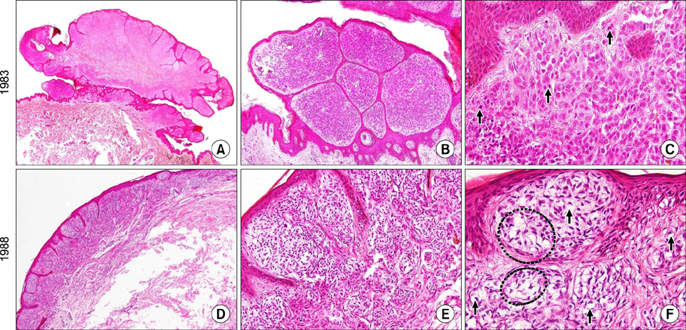
FIG. 1 Original slides dating back to 1983: the nevoid melanoma showed a verrucous growth pattern at scanning magnification (A, haematoxylin/eosin, ×1.25), peripheral densely cellular nodules with epidermal thinning at higher magnification (B, haematoxylin/eosin, ×10), and some mitotic figures pointed out by black arrows (C, haematoxylin/eosin, ×40). Original slides dating back to 1988: the signet-ring cell melanoma did not show a verrucous appearance (D, haematoxylin/eosin, ×4), it consisted of clear atypical cells (E, haematoxylin/eosin, ×20) with a signet-ring (F, arrows) or physalipherous (F, circles) morphology (F, haematoxylin/eosin, ×40).
Reference
-
1. Roncati L, Piscioli F, Pusiol T. Clinical application of the unifying concept of cutaneous melanoma. Chonnam Med J. 2017; 53:78–80.
Article2. Elder DE. Melanoma progression. Pathology. 2016; 48:147–154.
Article3. Lefevre M, Vergier B, Balme B, Thiebault R, Delaunay M, Thomas L, et al. Relevance of vertical growth pattern in thin level II cutaneous superficial spreading melanomas. Am J Surg Pathol. 2003; 27:717–724.
Article4. Gershenwald JE, Scolyer RA, Hess KR, Thompson JF, Long GV, Ross MI, et al. Melanoma of the skin. In : Amin MB, editor. AJCC cancer staging manual. 8th ed. Chicago: Springer;2017. p. 563–585.5. Breslow A. Thickness, cross-sectional areas and depth of invasion in the prognosis of cutaneous melanoma. Ann Surg. 1970; 172:902–908.
Article6. Clark WH Jr, From L, Bernardino EA, Mihm MC. The histogenesis and biologic behavior of primary human malignant melanomas of the skin. Cancer Res. 1969; 29:705–727.7. Büttner P, Garbe C, Bertz J, Burg G, d'Hoedt B, Drepper H, et al. Primary cutaneous melanoma. Optimized cutoff points of tumor thickness and importance of Clark's level for prognostic classification. Cancer. 1995; 75:2499–2506.
Article8. Balch CM, Soong SJ, Gershenwald JE, Thompson JF, Reintgen DS, Cascinelli N, et al. Prognostic factors analysis of 17,600 melanoma patients: validation of the American Joint Committee on Cancer melanoma staging system. J Clin Oncol. 2001; 19:3622–3634.
Article9. Roncati L, Pusiol T, Piscioli F. Thin melanoma: a generic term including four histological subtypes of cutaneous melanoma. Acta Dermatovenerol Croat. 2016; 24:169–174.10. Piscioli F, Pusiol T, Roncati L. Histopathological determination of thin melanomas at risk for metastasis. Melanoma Res. 2016; 26:635.
Article11. Bastian BC, Lazar A. Nevoid melanoma. In : Calonje E, Brenn T, Lazar A, McKee PH, editors. McKee's pathology of the skin: with clinical correlations. 4th ed. Edinburgh: Elsevier Saunders;2012. p. 1240.12. Magro CM, Crowson AN, Mihm MC. Unusual variants of malignant melanoma. Mod Pathol. 2006; 19:Suppl 2. S41–S70.
Article
