Effects of various cone-beam computed tomography settings on the detection of recurrent caries under restorations in extracted primary teeth
- Affiliations
-
- 1Department of Dentomaxillofacial Radiology, Faculty of Dentistry, Ankara University, Ankara, Turkey. dtkivo@yahoo.com
- 2Department of Biostatistics, Faculty of Veterinary Medicine, Ankara University, Ankara, Turkey.
- KMID: 2389891
- DOI: http://doi.org/10.5624/isd.2017.47.2.109
Abstract
- PURPOSE
The aim of this study was to assess the ex vivo diagnostic ability of 9 different cone-beam computed tomography (CBCT) settings in the detection of recurrent caries under amalgam restorations in primary teeth.
MATERIALS AND METHODS
Fifty-two primary teeth were used. Twenty-six teeth had dentine caries and 26 teeth did not have dentine caries. Black class II cavities were prepared and restored with amalgam. In the 26 carious teeth, recurrent caries were left under restorations. The other 26 intact teeth that did not have caries served as controls. Teeth were imaged using a 100×90-mm field of view and a 0.2-mm voxel size with 9 different CBCT settings. Four observers assessed the images using a 5-point scale. Kappa values were calculated to assess observer agreement. CBCT settings were compared with the gold standard using a receiver operating characteristic analysis. The area under the curve (AUC) values for each setting were compared using the chi-square test, with a significance level of α=.05.
RESULTS
Intraobserver kappa values ranged from 0.366 to 0.664 for observer 1, from 0.311 to 0.447 for observer 2, from 0.597 to 1.000 for observer 3, and from 0.869 to 1 for observer 4. Furthermore, interobserver kappa values among the observers ranged from 0.133 to 0.814 for the first reading and from 0.197 to 0.805 for the second reading. The highest AUC values were found for setting 5 (0.5916) and setting 3 (0.5886), and were not found to be statistically significant (P>.05).
CONCLUSION
Variations in tube voltage and tube current did not affect the detection of recurrent caries under amalgam restorations in primary teeth.
MeSH Terms
Figure
-

Fig. 1 Cross-sectional cone beam computed tomography images obtained by Planmeca Promax 3D Max. A. Setting with 60 kVp and 1 mA amalgam restoration with recurrent caries. B. Setting with 60 kVp and 6.3 mA amalgam restoration with beam hardening artifact. C. Setting with 60 kVp and 12.5 mA amalgam restoration without recurrent caries. D. Setting with 78 kVp and 1 mA amalgam restoration with recurrent caries. E. Setting with 78 kVp and 6.3 mA amalgam restoration with beam hardening artifact. F. Setting with 60 kVp and 12.5 mA amalgam restoration without recurrent caries. G. Setting with 96 kVp and 1 mA amalgam restoration with recurrent caries. H. Setting with 96 kVp and 6.3 mA amalgam restoration with beam hardening artifact. I. Setting with 96 kVp and 12.5 mA amalgam restoration without recurrent caries.
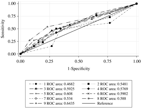
Fig. 2 Receiver operating characteristic curves for observer 3 for the first reading for all settings.
Reference
-
1. American Academy of Pediatric Dentistry. Guideline on caries-risk assessment and management for infants, children, and adolescents. Pediatr Dent. 2013; 35:E157–E164.2. Mjör IA, Toffenetti F. Secondary caries: a literature review with case reports. Quintessence Int. 2000; 31:165–179.3. Okida RC, Mandarino F, Sundfeld RH, de Alexandre RS, Sundefeld ML. In vitro-evaluation of secondary caries formation around restoration. Bull Tokyo Dent Coll. 2008; 49:121–128.
Article4. Ando M, González-Cabezas C, Isaacs RL, Eckert GJ, Stookey GK. Evaluation of several techniques for the detection of secondary caries adjacent to amalgam restorations. Caries Res. 2004; 38:350–356.
Article5. Murat S, Kamburoğlu K, Isayev A, Kurşun S, Yüksel S. Visibility of artificial buccal recurrent caries under restorations using different radiographic techniques. Oper Dent. 2013; 38:197–207.
Article6. Kamburoğlu K, Ilker Cebeci AR, Gröndahl HG. Effectiveness of limited cone-beam computed tomography in the detection of horizontal root fracture. Dent Traumatol. 2009; 25:256–261.
Article7. Mialhe FL, Pereira AC, Meneghim Mde C, Ambrosano GM, Pardi V. The relative diagnostic yields of clinical, FOTI and radiographic examinations for the detection of approximal caries in youngsters. Indian J Dent Res. 2009; 20:136–140.
Article8. Tveit AB, Espelid I. Class II amalgams: interobserver variations in replacement decisions and diagnosis of caries and crevices. Int Dent J. 1992; 42:12–18.9. Tyndall DA, Rathore S. Cone-beam CT diagnostic applications: caries, periodontal bone assessment, and endodontic applications. Dent Clin North Am. 2008; 52:825–841.
Article10. White SC. Cone-beam imaging in dentistry. Health Phys. 2008; 95:628–637.
Article11. Kamburoğlu K, Kurt H, Kolsuz E, Öztaş B, Tatar I, Çelik HH. Occlusal caries depth measurements obtained by five different imaging modalities. J Digit Imaging. 2011; 24:804–813.
Article12. Baltacıoĝlu İH, Eren H, Yavuz Y, Kamburoğlu K. Diagnostic accuracy of different display types in detection of recurrent caries under restorations by using CBCT. Dentomaxillofac Radiol. 2016; 45:20160099.
Article13. Kau CH, Bozic M, English J, Lee R, Bussa H, Ellis RK. Cone-beam computed tomography of the maxillofacial region-an update. Int J Med Robot. 2009; 5:366–380.
Article14. Svenson B, Welander U, Anneroth G, Söderfeldt B. Exposure parameters and their effects on diagnostic accuracy. Oral Surg Oral Med Oral Pathol. 1994; 78:544–550.
Article15. Sogur E, Baksı BG, Orhan K, Paksoy SC, Dogan S, Erdal YS, et al. Effect of tube potential and image receptor on the detection of natural proximal caries in primary teeth. Clin Oral Investig. 2011; 15:901–907.
Article16. Kaeppler G, Dietz K, Reinert S. Influence of tube potential setting and dose on the visibility of lesions in intraoral radiography. Dentomaxillofac Radiol. 2007; 36:75–79.
Article17. Landis JR, Koch GG. The measurement of observer agreement for categorical data. Biometrics. 1977; 33:159–174.
Article18. Yamaguchi K, Miyazaki M, Takamizawa T, Inage H, Moore BK. Effect of CPP-ACP paste on mechanical properties of bovine enamel as determined by an ultrasonic device. J Dent. 2006; 34:230–236.
Article19. Wilson PR, Beynon AD. Mineralization differences between human deciduous and permanent enamel measured by quantitative microradiography. Arch Oral Biol. 1989; 34:85–88.
Article20. Senel B, Kamburoglu K, Uçok O, Yüksel SP, Ozen T, Avsever H. Diagnostic accuracy of different imaging modalities in detection of proximal caries. Dentomaxillofac Radiol. 2010; 39:501–511.21. Kulczyk T, Dyszkiewicz Konwińska M, Owecka M, Krzyżostaniak J, Surdacka A. The influence of amalgam fillings on the detection of approximal caries by cone beam CT: in vitro study. Dentomaxillofac Radiol. 2014; 43:20130342.22. Safi Y, Hosseinpour S, Aziz A, Bamedi M, Malekashtari M, Vasegh Z. Effect of amperage and field of view on detection of vertical root fracture in teeth with intracanal posts. Iran Endod J. 2016; 11:202–207.23. Pinto MG, Rabelo KA, Sousa Melo SL, Campos PS, Oliveira LS, Bento PM, et al. Influence of exposure parameters on the detection of simulated root fractures in the presence of various intracanal materials. Int Endod J. 2017; 50:586–594.
Article24. Theodorakou C, Walker A, Horner K, Pauwels R, Bogaerts R, Jacobs R, et al. Estimation of paediatric organ and effective doses from dental cone beam CT using anthropomorphic phantoms. Br J Radiol. 2012; 85:153–160.
Article
- Full Text Links
-
- Actions
-
Cited
- CITED
-
- Close
- Share
-
- Similar articles
-
- A comparative study of cone-beam computed tomography and digital panoramic radiography for detecting pulp stones
- Current status of dental caries diagnosis using cone beam computed tomography
- Diagnostic performance of cone-beam computed tomography on detection of mechanically-created artificial secondary caries
- Evaluation of the relation between the pulp stones and direct restorations using cone beam computed tomography in a Turkish subpopulation
- Potential impact of metal crowns at varying distances from a carious lesion on its detection on cone-beam computed tomography scans with several protocols
