Treatment of Osteochondral Lesions of the Talus in Athletes
- Affiliations
-
- 1Department of Orthopedics, Soonchunhyang University Bucheon Hospital, Bucheon, Korea. brain0808@hanmail.net
- 2Department of Orthopedics, Veterans Health Service Medical Center, Seoul, Korea.
- KMID: 2389646
- DOI: http://doi.org/10.5763/kjsm.2017.35.2.77
Abstract
- The definition of osteochondral lesion of the talus (OLT) is any defect involving both the articular surface and the subchondral bone of the talus. Many of these lesions are associated with acute ankle injury. Although many classification schemes for OLT have been proposed, Berndt and Harty's 4-staging classification is most commonly used. Stage 4 lesions and symptomatic lesions under grade 3 are usually recommended to surgical treatment. The treatment approach for athletes should be more elaborate due to the need for an early return to play. Several different types of treatment are described for OLTs in athletes, including bone marrow stimulation, osteochondral autograft transfer system, and autogenous chondrocyte implantation. Osteochondral autograft transfer system shows good clinical outcome and has the advantages that could be applied to large defect and recurred lesions, however, it has some disadvantages in terms of the complications related with the donor site and the difficult approach to the medial lesions. Although autogenous chondrocyte implantation has been extensively applied for treating OLTs with successful clinical outcomes, it has some limitations that apply to athletes in terms of the 2-stage and complicated procedure and the insurance issues. Bone marrow stimulation being a simple and cost-effective procedure associated with a low complication rate and low postoperative pain has faster return to play and is recommended the first-line treatment for the OLTs of athletes.
Keyword
MeSH Terms
Figure
-
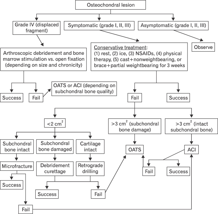
Fig. 1 The treatment algorithm for osteochondral lesion of the talus. NSAIDs: non-steroidal anti-inflammatory drugs, OATS: osteochondral autograft transfer system, ACI: autogenous chondrocyte implantation. Reproduced from Dragoni et al. Foot Ankle Int 2011;32:910-6, with permission of SAGE Publications 26).
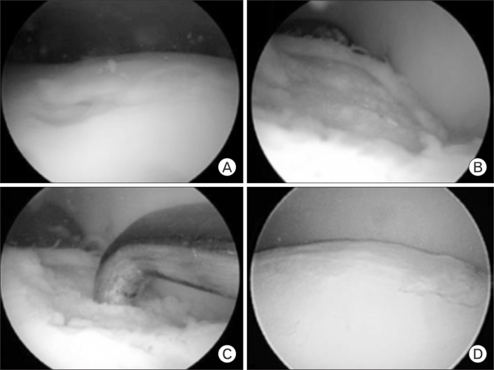
Fig. 2 Arthroscopic microfracture technique. (A) Arthroscopic view of the medial talar dome lesion. (B) The same lesion after curettage and (C) microfracture. (D) The healed lesion filling with fibrous cartilage after 2 years.

Fig. 3 The fluoroscopic images of osteochondral autograft transfer system. (A) The resection of the lateral malleolus was done during the approach to the lateral talar dome lesion. (B) The image shows plating of the lateral malleolus after the procedure.

Fig. 4 These pictures show second look arthroscopy after autologous chondrocyte implantation of osteochondral lesion of the talus. (A, B) Complete degree of defect repair and filling of the defect, (C, D) <50% incomplete degree of defect repair and filling of the defect. Reproduced from Lee et al. Scand J Med Sci Sports 2012;22:510-550).
Reference
-
1. Monro A. Part of the cartilage of the joint separated and ossified. Medical essays and observations. 2nd ed. Edinburgh: T and W Ruddimans;1737.2. Berndt AL, Harty M. Transchondral fractures (osteochondritis dissecans) of the talus. J Bone Joint Surg Am. 1959; 41:988–1020.3. O'Loughlin PF, Heyworth BE, Kennedy JG. Current concepts in the diagnosis and treatment of osteochondral lesions of the ankle. Am J Sports Med. 2010; 38:392–404.4. Draper SD, Fallat LM. Autogenous bone grafting for the treatment of talar dome lesions. J Foot Ankle Surg. 2000; 39:15–23.5. Leontaritis N, Hinojosa L, Panchbhavi VK. Arthroscopically detected intra-articular lesions associated with acute ankle fractures. J Bone Joint Surg Am. 2009; 91:333–339.6. Saxena A, Eakin C. Articular talar injuries in athletes: results of microfracture and autogenous bone graft. Am J Sports Med. 2007; 35:1680–1687.7. Aigner J, Tegeler J, Hutzler P, et al. Cartilage tissue engineering with novel nonwoven structured biomaterial based on hyaluronic acid benzyl ester. J Biomed Mater Res. 1998; 42:172–181.8. Angermann P, Jensen P. Osteochondritis dissecans of the talus: long-term results of surgical treatment. Foot Ankle. 1989; 10:161–163.9. Buckwalter JA, Lohmander S. Operative treatment of osteoarthrosis: current practice and future development. J Bone Joint Surg Am. 1994; 76:1405–1418.10. Navid DO, Myerson MS. Approach alternatives for treatment of osteochondral lesions of the talus. Foot Ankle Clin. 2002; 7:635–649.11. Verhagen RA, Struijs PA, Bossuyt PM, van Dijk CN. Systematic review of treatment strategies for osteochondral defects of the talar dome. Foot Ankle Clin. 2003; 8:233–242.12. Flick AB, Gould N. Osteochondritis dissecans of the talus (transchondral fractures of the talus): review of the literature and new surgical approach for medial dome lesions. Foot Ankle. 1985; 5:165–185.13. Aspenberg P, Van der Vis H. Migration, particles, and fluid pressure: a discussion of causes of prosthetic loosening. Clin Orthop Relat Res. 1998; (352):75–80.14. Looze CA, Capo J, Ryan MK, et al. Evaluation and management of osteochondral lesions of the talus. Cartilage. 2017; 8:19–30.15. Frenkel SR, Di Cesare PE. Degradation and repair of articular cartilage. Front Biosci. 1999; 4:D671–D685.16. Elias I, Jung JW, Raikin SM, Schweitzer MW, Carrino JA, Morrison WB. Osteochondral lesions of the talus: change in MRI findings over time in talar lesions without operative intervention and implications for staging systems. Foot Ankle Int. 2006; 27:157–166.17. Astrand J, Skripitz R, Skoglund B, Aspenberg P. A rat model for testing pharmacologic treatments of pressure-related bone loss. Clin Orthop Relat Res. 2003; (409):296–305.18. Van der Vis HM, Aspenberg P, Marti RK, Tigchelaar W, Van Noorden CJ. Fluid pressure causes bone resorption in a rabbit model of prosthetic loosening. Clin Orthop Relat Res. 1998; (350):201–208.19. Goddard NJ, Gosling PT. Intra-articular fluid pressure and pain in osteoarthritis of the hip. J Bone Joint Surg Br. 1988; 70:52–55.20. Saxler G, Loer F, Skumavc M, Pfortner J, Hanesch U. Localization of SP- and CGRP-immunopositive nerve fibers in the hip joint of patients with painful osteoarthritis and of patients with painless failed total hip arthroplasties. Eur J Pain. 2007; 11:67–74.21. Pritsch M, Horoshovski H, Farine I. Arthroscopic treatment of osteochondral lesions of the talus. J Bone Joint Surg Am. 1986; 68:862–865.22. Hepple S, Winson IG, Glew D. Osteochondral lesions of the talus: a revised classification. Foot Ankle Int. 1999; 20:789–793.23. Dipaola JD, Nelson DW, Colville MR. Characterizing osteochondral lesions by magnetic resonance imaging. Arthroscopy. 1991; 7:101–104.24. Mintz DN, Tashjian GS, Connell DA, Deland JT, O'Malley M, Potter HG. Osteochondral lesions of the talus: a new magnetic resonance grading system with arthroscopic correlation. Arthroscopy. 2003; 19:353–359.25. Ferkel RD, Zanotti RM, Komenda GA, et al. Arthroscopic treatment of chronic osteochondral lesions of the talus: long-term results. Am J Sports Med. 2008; 36:1750–1762.26. Dragoni M, Bonasia DE, Amendola A. Osteochondral talar allograft for large osteochondral defects: technique tip. Foot Ankle Int. 2011; 32:910–916.27. Amendola A, Panarella L. Osteochondral lesions: medial versus lateral, persistent pain, cartilage restoration options and indications. Foot Ankle Clin. 2009; 14:215–227.28. Hannon CP, Smyth NA, Murawski CD, et al. Osteochondral lesions of the talus: aspects of current management. Bone Joint J. 2014; 96-B:164–171.29. Klammer G, Maquieira GJ, Spahn S, Vigfusson V, Zanetti M, Espinosa N. Natural history of nonoperatively treated osteochondral lesions of the talus. Foot Ankle Int. 2015; 36:24–31.30. Lee KT, Young KW, Lee YK, Park SY, Jang MS. Results in conservative treatment of osteochondral lesion of talus. J Korean Foot Ankle Soc. 2008; 12:145–149.31. Murawski CD, Kennedy JG. Operative treatment of osteochondral lesions of the talus. J Bone Joint Surg Am. 2013; 95:1045–1054.32. Kennedy JG, Murawski CD. The treatment of osteochondral lesions of the talus with autologous osteochondral transplantation and bone marrow aspirate concentrate: surgical technique. Cartilage. 2011; 2:327–336.33. Zengerink M, Struijs PA, Tol JL, van Dijk CN. Treatment of osteochondral lesions of the talus: a systematic review. Knee Surg Sports Traumatol Arthrosc. 2010; 18:238–246.34. Porter DA, Schon LC. Baxter's the foot and ankle in sport. 2nd ed. Philadelphia: Mosby Elsevier;2008.35. Bae DK, Yoon KH, Song SJ. Cartilage healing after microfracture in osteoarthritic knees. Arthroscopy. 2006; 22:367–374.36. Mithoefer K, Hambly K, Logerstedt D, Ricci M, Silvers H, Della Villa S. Current concepts for rehabilitation and return to sport after knee articular cartilage repair in the athlete. J Orthop Sports Phys Ther. 2012; 42:254–273.37. Lee DH, Lee KB, Jung ST, Seon JK, Kim MS, Sung IH. Comparison of early versus delayed weightbearing outcomes after microfracture for small to midsized osteochondral lesions of the talus. Am J Sports Med. 2012; 40:2023–2028.38. Choi WJ, Park KK, Kim BS, Lee JW. Osteochondral lesion of the talus: is there a critical defect size for poor outcome? Am J Sports Med. 2009; 37:1974–1980.39. Chuckpaiwong B, Berkson EM, Theodore GH. Microfracture for osteochondral lesions of the ankle: outcome analysis and outcome predictors of 105 cases. Arthroscopy. 2008; 24:106–112.40. Csonge L, Bravo D, Newman-Gage H, et al. Banking of osteochondral allografts. Part II: preservation of chondrocyte viability during long-term storage. Cell Tissue Bank. 2002; 3:161–168.41. Schachar NS, McGann LE. Investigations of low-temperature storage of articular cartilage for transplantation. Clin Orthop Relat Res. 1986; (208):146–150.42. Tomford WW, Duff GP, Mankin HJ. Experimental freeze-preservation of chondrocytes. Clin Orthop Relat Res. 1985; (197):11–14.43. Imhoff AB, Paul J, Ottinger B, et al. Osteochondral transplantation of the talus: long-term clinical and magnetic resonance imaging evaluation. Am J Sports Med. 2011; 39:1487–1493.44. Scranton PE Jr, Frey CC, Feder KS. Outcome of osteochondral autograft transplantation for type-V cystic osteochondral lesions of the talus. J Bone Joint Surg Br. 2006; 88:614–619.45. Kreuz PC, Steinwachs M, Erggelet C, Lahm A, Henle P, Niemeyer P. Mosaicplasty with autogenous talar autograft for osteochondral lesions of the talus after failed primary arthroscopic management: a prospective study with a 4-year follow-up. Am J Sports Med. 2006; 34:55–63.46. Sammarco GJ, Makwana NK. Treatment of talar osteochondral lesions using local osteochondral graft. Foot Ankle Int. 2002; 23:693–698.47. LaPrade RF, Botker JC. Donor-site morbidity after osteochondral autograft transfer procedures. Arthroscopy. 2004; 20:e69–e73.48. Peters PG, Parks BG, Schon LC. Anterior distal tibia plafondplasty for exposure of the talar dome. Foot Ankle Int. 2012; 33:231–235.49. Young KW, Deland JT, Lee KT, Lee YK. Medial approaches to osteochondral lesion of the talus without medial malleolar osteotomy. Knee Surg Sports Traumatol Arthrosc. 2010; 18:634–637.50. Lee KT, Lee YK, Young KW, Park SY, Kim JS. Factors influencing result of autologous chondrocyte implantation in osteochondral lesion of the talus using second look arthroscopy. Scand J Med Sci Sports. 2012; 22:510–515.51. Paul J, Sagstetter M, Lammle L, et al. Sports activity after osteochondral transplantation of the talus. Am J Sports Med. 2012; 40:870–874.52. Tegner Y, Lysholm J. Rating systems in the evaluation of knee ligament injuries. Clin Orthop Relat Res. 1985; (198):43–49.53. Marx RG, Stump TJ, Jones EC, Wickiewicz TL, Warren RF. Development and evaluation of an activity rating scale for disorders of the knee. Am J Sports Med. 2001; 29:213–218.54. Kwak SK, Kern BS, Ferkel RD, Chan KW, Kasraeian S, Applegate GR. Autologous chondrocyte implantation of the ankle: 2- to 10-year results. Am J Sports Med. 2014; 42:2156–2164.55. Giannini S, Buda R, Grigolo B, Vannini F. Autologous chondrocyte transplantation in osteochondral lesions of the ankle joint. Foot Ankle Int. 2001; 22:513–517.56. Lee KT, Choi YS, Lee YK, Kim JS, Young KW, Kim JH. Comparison of MRI and arthroscopy after autologous chondrocyte implantation in patients with osteochondral lesion of the talus. Orthopedics. 2010; 33.57. Lee KT, Choi YS, Lee YK, Cha SD, Koo HM. Comparison of MRI and arthroscopy in modified MOCART scoring system after autologous chondrocyte implantation for osteochondral lesion of the talus. Orthopedics. 2011; 34:e356–e362.
- Full Text Links
-
- Actions
-
Cited
- CITED
-
- Close
- Share
-
- Similar articles
-
- Simultaneously Occurred Medial and Lateral Osteochondral Lesions of the Talus
- Autologous Osteochondral Transplantation as a Secondary Procedure after Failed Microfracture for Osteochondral Lesion of Talus
- Osteochondral Lesions of the Talus: Autologous Osteochondral Transplantation
- Current Updates in Treatment of Osteochondral Lesions of the Talus
- Osteochondral Lesions of the Talus
