Lipoma Compressing the Sciatic Nerve in a Patient With Suspicious Central Post-stroke Pain
- Affiliations
-
- 1Department of Rehabilitation Medicine, College of Medicine, The Catholic University of Korea, Seoul, Korea.
- 2Department of Rehabilitation Medicine, Dongsan Medical Center, Keimyung University School of Medicine, Daegu, Korea. ymchoi@dsmc.or.kr
- KMID: 2389466
- DOI: http://doi.org/10.5535/arm.2017.41.3.488
Abstract
- Lipomas are mostly located in the subcutaneous tissues and rarely cause symptoms. Occasionally, peripheral nerve compression by lipomas is reported. We describe a case of a 59-year-old man with a left-middle cerebral artery infarction who was newly diagnosed as right basal ganglia and thalamic intracranial hemorrhage. He had neuropathic pain in the left arm and leg that was suspected to be central post-stroke pain. The administration of pain medication brought only temporary symptom relief. Nerve conduction and electromyography studies revealed left L5 radiculopathy and he showed a positive "˜sign of the buttock' in the left hip. Left-hip magnetic resonance imaging revealed an intermuscular lipoma compressing the sciatic nerve. After surgery, the range of motion in the left hip joint was significantly increased, and the patient's pain was relieved.
Keyword
MeSH Terms
Figure
-
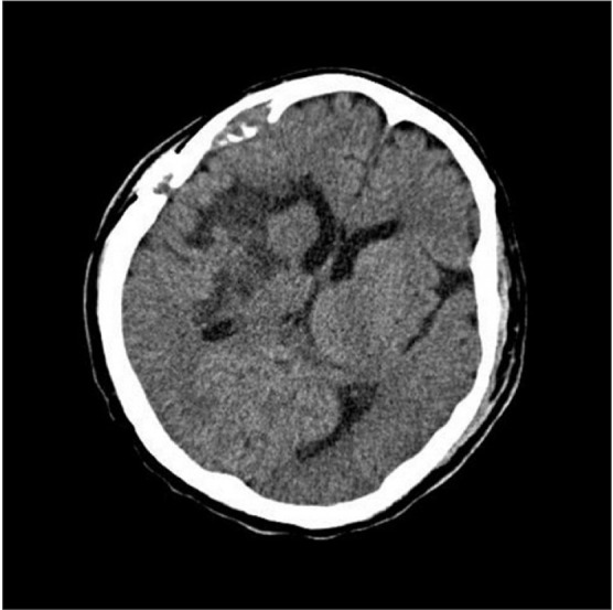
Fig. 1 Brain-computed tomography upon first admission showed encephalomalacic change in the right basal ganglia and thalamus caused by an intracranial hemorrhage.
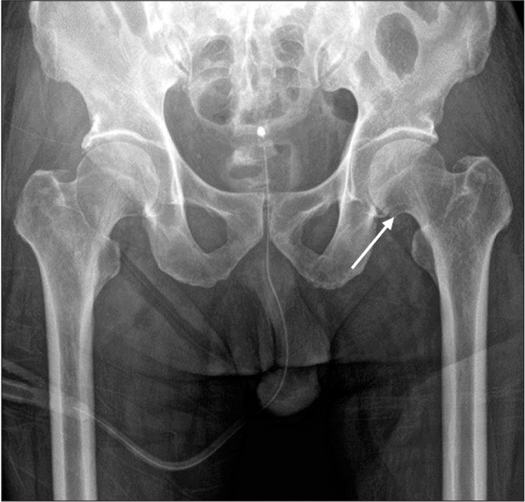
Fig. 2 Hip radiography revealed an osteophyte (arrow) in the left femoral head.
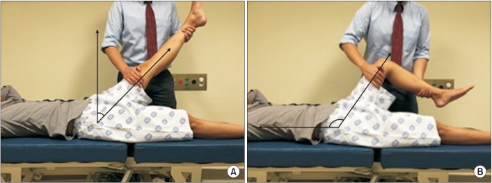
Fig. 3 The sign of the buttock: passive hip flexion with bent knee (B) is more limited and/or painful than straight leg raising (A).
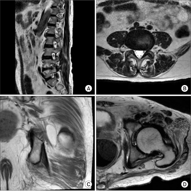
Fig. 4 T2-weighted magnetic resonance imaging (MRI) of the lumbosacral area showed diffuse bulging of the L4-5 disc. Sagittal view (A) and axial view (B) at L4-5 level. T1-weighted MRI of the left hip showed deep soft tissue in the ischiofemoral space, measuring about 4.0 cm×1.1 cm×2.2 cm around the sciatic nerve. Coronal view (C) and axial view (D).
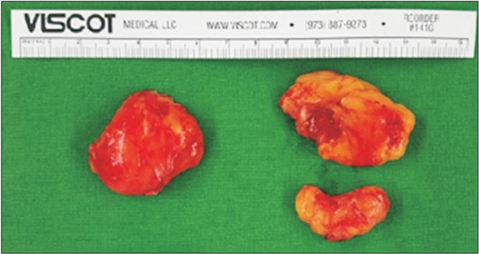
Fig. 5 Gross pathology of lipoma in the left hip lesion, a 4 cm×4 cm×2 cm encapsulated fatty mass between the gluteus medius and maximus that adhered to the sciatic nerve.
Reference
-
1. Kaeser MA, Smith LW, Kettner NW. A case report of an intermuscular lipoma: presentation, pathophysiology, differential diagnosis. J Chiropr Med. 2010; 9:127–131. PMID: 22027035.
Article2. Erhard RE, Egloff BP. Patient with metastatic adenocarcinoma imitating lumbar herniated nucleus pulposis. J Manipulative Physiol Ther. 2004; 27:569–573. PMID: 15614244.
Article3. Klit H, Finnerup NB, Jensen TS. Central post-stroke pain: clinical characteristics, pathophysiology, and management. Lancet Neurol. 2009; 8:857–868. PMID: 19679277.
Article4. Bugnicourt JM, Peltier J, Merle PE, Le Gars D. Acute peripheral nerve compression by a lipoma mimicking stroke. Clin Neurol Neurosurg. 2009; 111:395–396. PMID: 19100680.
Article5. Botwin KP, Shah CP, Zak PJ. Sciatic neuropathy secondary to infiltrating intermuscular lipoma of the thigh. Am J Phys Med Rehabil. 2001; 80:754–758. PMID: 11562557.
Article6. Kumar B, Kalita J, Kumar G, Misra UK. Central poststroke pain: a review of pathophysiology and treatment. Anesth Analg. 2009; 108:1645–1657. PMID: 19372350.
Article7. Greenwood MJ, Erhard RE, Jones DL. Differential diagnosis of the hip vs. lumbar spine: five case reports. J Orthop Sports Phys Ther. 1998; 27:308–315. PMID: 9549715.
Article8. Kong KH, Woon VC, Yang SY. Prevalence of chronic pain and its impact on health-related quality of life in stroke survivors. Arch Phys Med Rehabil. 2004; 85:35–40. PMID: 14970965.
- Full Text Links
-
- Actions
-
Cited
- CITED
-
- Close
- Share
-
- Similar articles
-
- Piriformis Syndrome (Sciatic Nerve Entrapment) Associated With Type C Sciatic Nerve Variation: A Report of Two Cases and Literature Review
- Ultrasound-Guided Sciatic Nerve Block for the Treatment of Radiation Therapy Induced Sciatic Neuritis: A case report
- Lipoma Causing Glossopharyngeal Neuralgia: A Case Report and Review of Literature
- Sural Nerve Graftafter Resection of a Schwannoma in the Sciatic Nerve : A Case Report
- Sciatic Neuropathy after Intramuscular Injection at a Site Remote from the Nerve
