Diagnosis of Ilioinguinal Nerve Injury Based on Electromyography and Ultrasonography: A Case Report
- Affiliations
-
- 1Department of Rehabilitation Medicine, Hallym University Kangdong Sacred Heart Hospital, Hallym University College of Medicine, Seoul, Korea. skyler02@hallym.or.kr
- KMID: 2389418
- DOI: http://doi.org/10.5535/arm.2017.41.4.705
Abstract
- Being located in the hypogastric area, the ilioinguinal nerve, together with iliohypogastric nerve, can be damaged during lower abdominal surgeries. Conventionally, the diagnosis of ilioinguinal neuropathy relies on clinical assessments, and standardized diagnostic methods have not been established as of yet. We hereby report the case of young man who presented ilioinguinal neuralgia with symptoms of burning pain in the right groin and scrotum shortly after receiving inguinal herniorrhaphy. To raise the diagnostic certainty, we used a real-time ultrasonography (US) to guide a monopolar electromyography needle to the ilioinguinal nerve, and then performed a motor conduction study. A subsequent US-guided ilioinguinal nerve block resulted in complete resolution of the patient's neuralgic symptoms.
MeSH Terms
Figure
-

Fig. 1 Herniorrhaphy site of the patient (white arrow).

Fig. 2 Ilioinguinal motor conduction study. (A) Ultrasound image of the ilioinguinal nerve and monopolar needle between the internal abdominal oblique muscle and transverses abdominis muscle (red arrow, tip of monopolar needle; yellow arrow, ilioinguinal nerve; EO, external abdominal oblique muscle; IO, internal abdominal oblique muscle; TA, transversus abdominis muscle). (B) Demonstration of conduction study (A, needle electrode; B, probe; C, recording electrode; D, reference electrode; E, ground electrode).

Fig. 3 Motor conduction study of ilioinguinal nerve. (A) Compound muscle action potential (CMAP) of left internal abdominal oblique muscle (latency, 4.8 ms; amplitude, 0.9 mV). (B) CMAP of right internal abdominal oblique muscle (no response).
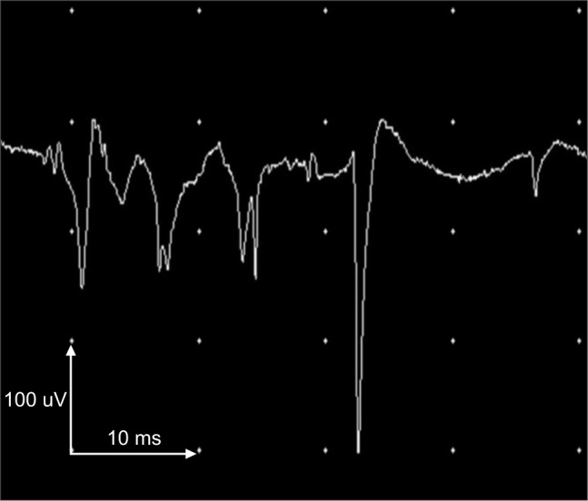
Fig. 4 Abnormal spontaneous activities in needle electromyography at the most caudal segment of the right internal abdominal oblique muscle.
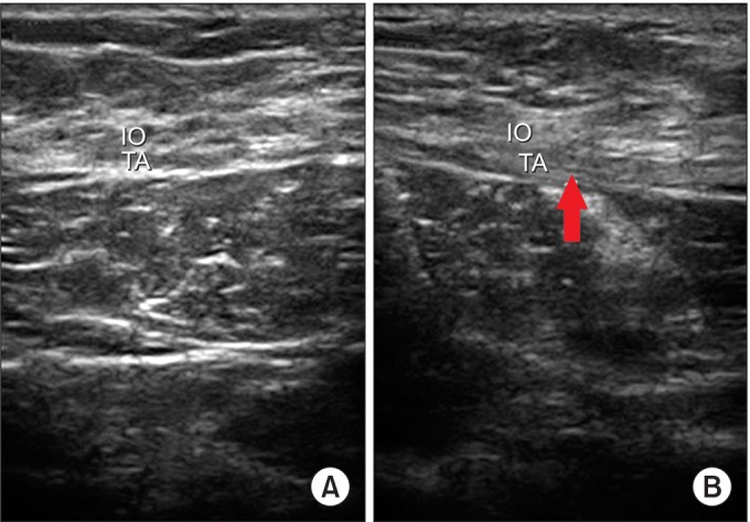
Fig. 5 Ultrasonography of lower abdominal muscle. (A) Left side (normal), (B) right side (red arrow, diffuse fibrotic change of muscles). IO, internal abdominal oblique muscle; TA, transversus abdominis muscle.
Reference
-
1. Rozen WM, Tran TM, Ashton MW, Barrington MJ, Ivanusic JJ, Taylor GI. Refining the course of the thoracolumbar nerves: a new understanding of the innervation of the anterior abdominal wall. Clin Anat. 2008; 21:325–333. PMID: 18428988.
Article2. ter Meulen BC, Peters EW, Wijsmuller A, Kropman RF, Mosch A, Tavy DL. Acute scrotal pain from idiopathic ilioinguinal neuropathy: diagnosis and treatment with EMG-guided nerve block. Clin Neurol Neurosurg. 2007; 109:535–537. PMID: 17481807.
Article3. Ellis RJ, Geisse H, Holub BA, Swenson MR. Ilioinguinal nerve conduction. Muscle Nerve. 1992; 10:1194.4. Karakayali F, Karatas M, Ozcelik U, Ekici Y, Basaran O, Moray G, et al. Influence of synthetic mesh on ilioinguinal nerve motor conduction and chronic groin pain after inguinal herniorrhaphy: a prospective randomized clinical study. Int Surg. 2007; 92:344–350. PMID: 18402129.
- Full Text Links
-
- Actions
-
Cited
- CITED
-
- Close
- Share
-
- Similar articles
-
- Origin and branching pattern of the iliohypogastric and ilioinguinal nerves and their exits in relation to the psoas major muscle: a cadaveric study
- Fracture of the Distal Radius with Ulnar Nerve Palsy
- Diagnostic Usefulness of Neuromuscular Ultrasound in Anatomical Localization of Peripheral Nerve Injury: Detailed Lesion Localization Using Neuromuscular Ultrasound in a Patient with Traumatic Ulnar Nerve Injury at the Hand
- Tourniquet Palsy of Median, Ulnar and Radial Nerves after Axillary Nerve Block: A case report
- Ilioinguinal and Iliohypogastric Nerve Block for Neuropathic Pain Following the Laparoscopic Surgery
