Primary Cutaneous Nocardiosis Caused by Nocardia takedensis
- Affiliations
-
- 1Department of Dermatology, Maryknoll Medical Center, Busan, Korea. heartthrob80@naver.com
- 2Department of Laboratory Medicine, Maryknoll Medical Center, Busan, Korea.
- 3Department of Dermatology, Kosin University College of Medicine, Busan, Korea.
- KMID: 2388946
- DOI: http://doi.org/10.5021/ad.2017.29.4.471
Abstract
- Nocardia species are aerobic, gram-positive, filamentous, partially acid-fast actinomycetes which are found worldwide in soil and decaying organic plant matter. When they infect human beings, they generally enter through the respiratory tract and then disseminate systemically. Rarely has a primary infection occurred as the result of direct inoculation. Isolation of Nocardia from clinical specimens and identification of species are difficult. But, with the introduction of new genetic technologies, reports of novel species of Nocardia have increased. We describe a case of cutaneous nocardiosis caused by Nocardia takedensis in an 87-year-old woman who was diagnosed by bacterial culture and 16S ribosomal RNA sequencing. N. takedensis has been described as a new species. This report describes the first clinical isolate of N. takedensis from a skin specimen in Korea.
Keyword
MeSH Terms
Figure
-

Fig. 1 Cutaneous nocardiosis from Nocardia takedensis infection. The left lower arm shows erythematous and edematous plaque, multiple ulcerations, abscesses, and black crusts. (A) Pretreatment lateral side, (B) pretreatment medial side, (C) posttreatment lateral side, (D) posttreatment medial side.
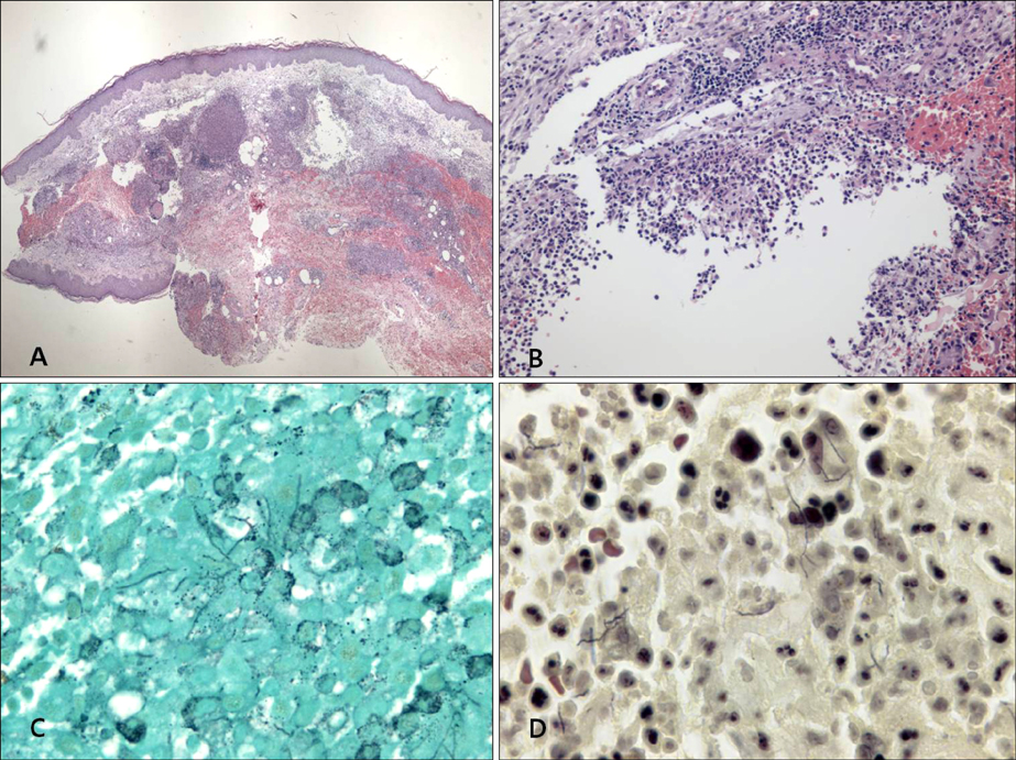
Fig. 2 Histopathologic findings. (A) Intense edema in the upper dermis and dense multinodular pandermal and subcutaneous neutrophilic infiltration (H&E, ×40). (B) Dense inflammatory infiltrates, with abscess formation, in the dermis (H&E, ×200). (C) Fine, filamentous branching bacilli (Grocott-Gomori's methenamine silver [GMS] stain, ×1,000). (D) Gram-positive rods with long, sinuous branches (Gram stain, ×1,000).

Fig. 3 Colony culture. (A) Ziehl-Neelsen stain revealing a partially positive result on cultured colony (Ziehl-Neelsen, ×1,000). (B) Chalky-orange-colored colonies formed on a blood agar plate incubated at 35℃ for 2 weeks.
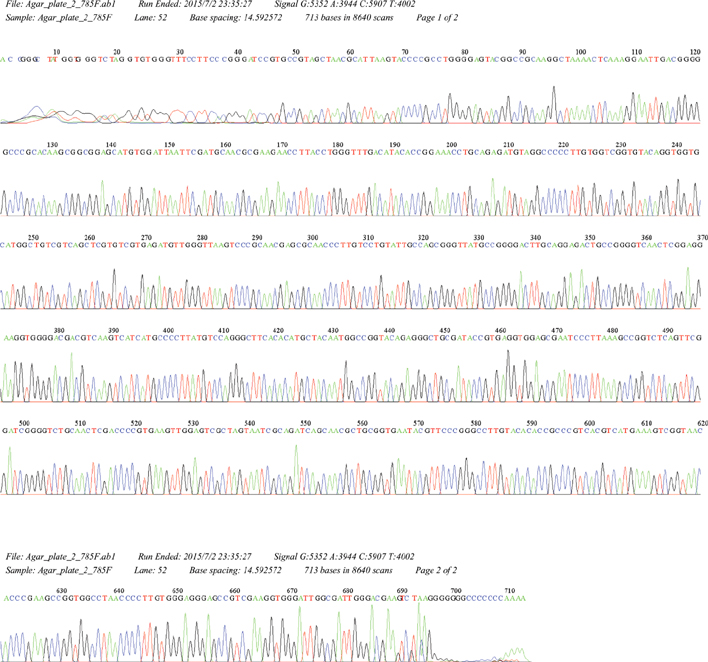
Fig. 4 16S ribosomal RNA (rRNA) gene sequencing. DNA was extracted from a cultured colony for polymerase chain reaction amplification and sequencing of both strands of the 16S rRNA gene. The sequence data confirmed the presence of Nocardia takedensis.
Reference
-
1. Brown-Elliott BA, Brown JM, Conville PS, Wallace RJ Jr. Clinical and laboratory features of the Nocardia spp. based on current molecular taxonomy. Clin Microbiol Rev. 2006; 19:259–282.
Article2. Saubolle MA, Sussland D. Nocardiosis: review of clinical and laboratory experience. J Clin Microbiol. 2003; 41:4497–4501.3. Lerner PI. Nocardiosis. Clin Infect Dis. 1996; 22:891–903.
Article4. Yamamura H, Hayakawa M, Nakagawa Y, Tamura T, Kohno T, Komatsu F, et al. Nocardia takedensis sp. nov., isolated from moat sediment and scumming activated sludge. Int J Syst Evol Microbiol. 2005; 55:433–436.
Article5. Japan MLJVTGJ. Takeda shrine. 2007-2012 [Internet]. MustLoveJapan;2014 Oct 29. Available from: http://www.mustlovejapan.com/subject/takeda_jinja/.6. Wilson JW. Nocardiosis: updates and clinical overview. Mayo Clin Proc. 2012; 87:403–407.
Article7. Chung E, Pulitzer MP, Papadopoulos EB, Papanicolaou GA, Babady NE, Marchetti MA. Lympahngitic papules caused by Nocardia takedensis. JAAD Case Rep. 2015; 1:126–128.8. Liu WL, Lai CC, Ko WC, Chen YH, Tang HJ, Huang YL, et al. Clinical and microbiological characteristics of infections caused by various Nocardia species in Taiwan: a multicenter study from 1998 to 2010. Eur J Clin Microbiol Infect Dis. 2011; 30:1341–1347.
Article9. Kresch-Tronik NS, Carrillo-Casas EM, Arenas R, Atoche C, Del Río-Ávila C, Ochoa-Carrera LA, et al. First case of mycetoma associated with Nocardia takedensis. J Dermatol. 2013; 40:135–136.
Article10. Mathur S, Sood R, Aron M, Iyer VK, Verma K. Cytologic diagnosis of pulmonary nocardiosis: a report of 3 cases. Acta Cytol. 2005; 49:567–570.11. Betrán A, Rezusta A, Lezcano MA, Villuendas MC, Revillo MJ, Boiron P, et al. First Spanish case of nocardiosis caused by Nocardia takedensis. J Clin Microbiol. 2009; 47:1918–1919.12. In SG, Han SH, Shin JH, Choi GS, Chung MH. A case of disseminated nocardiosis secondary to the skin nodules in an elderly woman. Ann Dermatol. 2008; 20:82–85.
Article13. Uhde KB, Pathak S, McCullum I Jr, Jannat-Khah DP, Shadomy SV, Dykewicz CA, et al. Antimicrobial-resistant nocardia isolates, United States, 1995-2004. Clin Infect Dis. 2010; 51:1445–1448.
Article
- Full Text Links
-
- Actions
-
Cited
- CITED
-
- Close
- Share
-
- Similar articles
-
- Primary Cutaneous Nocardiosis Caused by Nocardia niigatensis
- Primary Cutaneous Nocardiosis Caused by Nocardia farcinica
- Primary Cutaneous Nocardiosis Caused by Nocardia brasiliensis
- A Case of Primary Cutaneous Sporotrichoid Nocardiosis Caused by Nocardia asteroides
- Nocardia asteroides complex Isolated from Cerebrospinal Fluid and Surgical Wound Site: Three Case Reports
