Plasma Cell Cheilitis: A Clinicopathological and Immunohistochemical Study of 13 Cases
- Affiliations
-
- 1Department of Dermatology, Kangdong Sacred Heart Hospital, Hallym University College of Medicine, Seoul, Korea. drkimss@naver.com
- 2Department of Dermatology, Hallym University Sacred Heart Hospital, Hallym University College of Medicine, Anyang, Korea.
- KMID: 2388903
- DOI: http://doi.org/10.5021/ad.2017.29.5.536
Abstract
- BACKGROUND
Plasma cell cheilitis is an unusual benign plasma cell proliferative disease of an unknown etiology that typically presents on the lip.
OBJECTIVE
The aim of this study was to investigate the clinicopathological characteristics of 13 cases of plasma cell cheilitis.
METHODS
The present study investigated the clinical manifestations, treatment modalities, and outcome of 13 patients diagnosed with plasma cell cheilitis from 2011 to 2016 at Kangdong Sacred Heart Hospital and Hallym University Sacred Heart Hospital. Biopsy specimens of the all cases were evaluated using conventional hematoxylin and eosin staining with kappa and lambda immunoglobulin light chain immunohistochemistry.
RESULTS
The age of the patients ranged from 39 to 86 years (mean, 64.7 years), with male predominance. Histopathologically, 61.5% and 38.5% of patients showed band-like and pan dermal plasmacytic infiltrates, respectively. Eosinophilic infiltration was noted in 69.2% of patients. All cases showed both kappa and lambda immunoglobulin light chain reactivities, and kappa predominance was confirmed in 9 patients (69.2%). A majority of the patients was treated with local therapy, such as intralesional steroid injection with topical tacrolimus. Among the 13 patients, plasma cell cheilitis completely resolved, partially resolved, and recurred in 3 (23.1%), 5 (38.5%), and 5 patients (38.5%), respectively.
CONCLUSION
Plasma cell cheilitis presented as erosive edematous circumscribed patches or plaques affecting mainly the lower lip of elderly male patients. The majority of histopathology cases showed characteristic plasma cell aggregation on the upper dermis that was immunopositive for immunoglobulin light chain, with kappa predominance.
Keyword
MeSH Terms
Figure
-
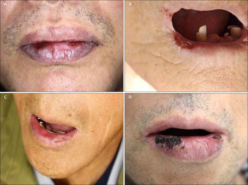
Fig. 1 (A) The lower lip shows a diffuse dark blue-grayish to erythematous patch with several fissures and hemorrhagic crusts (patient 3). (B) Painful ulcerative lesion is noted on the left lower angle of the lip on a patient who used dentures (patient 9). (C) Prominent edematous erosive lower lip mucosa is shown (patient 10). (D) Multiple well-demarcated erythematous patches with crust formation and marked swelling on the lower lip are identified (patient 6).
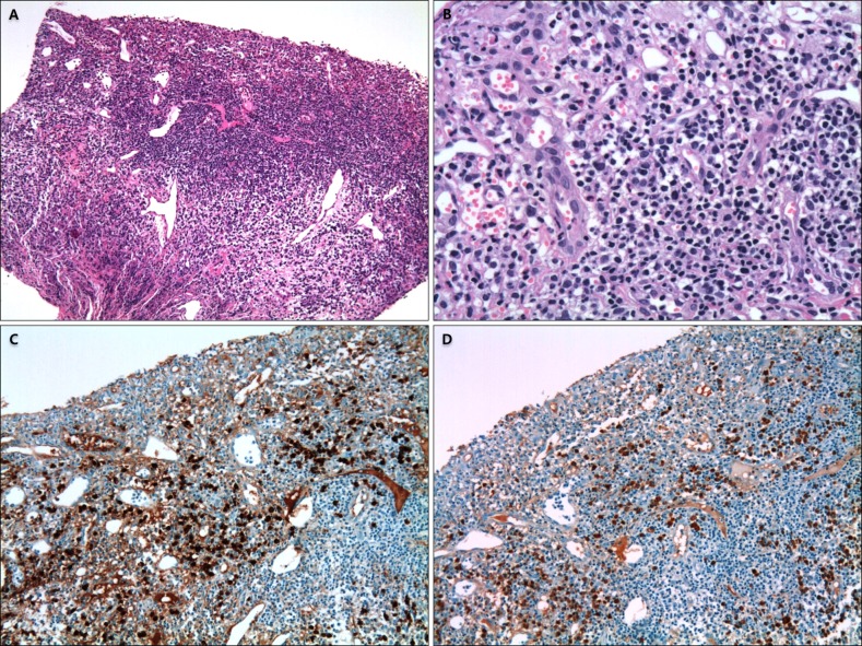
Fig. 2 This specimen from the lip shows an epidermal detachment with a dense, band-like infiltrate of plasma cells in the upper dermis. (A) Eosinophilic infiltration within the dermis is also present (H&E, ×100). (B) A high power view shows a monomorphic population of mature plasma cells without cellular atypia, pleomorphic figures, and mitotic activity (H&E, ×400). (C) A diffuse strong positivity for kappa light chain is noted in the plasma cell infiltrates (kappa chain, ×200). (D) Few plasma cells are positive for lambda light chain restriction (lambda chain, ×200).
Reference
-
1. Rocha N, Mota F, Horta M, Lima O, Massa A, Sanches M. Plasma cell cheilitis. J Eur Acad Dermatol Venereol. 2004; 18:96–98. PMID: 14678543.
Article2. Farrier JN, Perkins CS. Plasma cell cheilitis. Br J Oral Maxillofac Surg. 2008; 46:679–680. PMID: 18439733.
Article3. Baughman RD, Berger P, Pringle WM. Plasma cell cheilitis. Arch Dermatol. 1974; 110:725–726. PMID: 4422015.
Article4. Solomon LW, Wein RO, Rosenwald I, Laver N. Plasma cell mucositis of the oral cavity: report of a case and review of the literature. Oral Surg Oral Med Oral Pathol Oral Radiol Endod. 2008; 106:853–860. PMID: 18926737.
Article5. Smith ME, Crighton AJ, Chisholm DM, Mountain RE. Plasma cell mucositis: a review and case report. J Oral Pathol Med. 1999; 28:183–186. PMID: 10235373.
Article6. Yang JH, Lee UH, Jang SJ, Choi JC. Plasma cell cheilitis treated with intralesional injection of corticosteroids. J Dermatol. 2005; 32:987–990. PMID: 16471463.
Article7. Aiba S, Tagami H. Immunoglobulin-producing cells in plasma cell orificial mucositis. J Cutan Pathol. 1989; 16:207–210. PMID: 2677069.
Article8. Tamaki K, Osada A, Tsukamoto K, Ohtake N, Furue M. Treatment of plasma cell cheilitis with griseofulvin. J Am Acad Dermatol. 1994; 30:789–790. PMID: 8176022.
Article9. Choi JW, Choi M, Cho KH. Successful treatment of plasma cell cheilitis with topical calcineurin inhibitors. J Dermatol. 2009; 36:669–671. PMID: 19958456.
Article10. Hanami Y, Motoki Y, Yamamoto T. Successful treatment of plasma cell cheilitis with topical tacrolimus: report of two cases. Dermatol Online J. 2011; 17:6.
Article11. Jin SP, Cho KH, Huh CH. Plasma cell cheilitis, successfully treated with topical 0.03% tacrolimus ointment. J Dermatolog Treat. 2010; 21:130–132. PMID: 19707925.
Article12. Tseng JT, Cheng CJ, Lee WR, Wang KH. Plasma-cell cheilitis: successful treatment with intralesional injections of corticosteroids. Clin Exp Dermatol. 2009; 34:174–177. PMID: 18795938.
Article13. Yoshimura K, Nakano S, Tsuruta D, Ohata C, Hashimoto T. Successful treatment with 308-nm monochromatic excimer light and subsequent tacrolimus 0.03% ointment in refractory plasma cell cheilitis. J Dermatol. 2013; 40:471–474. PMID: 23621817.
Article14. Noorily AD. Plasma cell orificial mucositis. Otolaryngol Head Neck Surg. 1997; 116:416–417. PMID: 9121804.
Article15. Choe HC, Park HJ, Oh ST, Park CJ, Byun DG, Cho BK. Clinicopathologic study of 8 patients with plasma cell cheilitis. Korean J Dermatol. 2003; 41:174–178.16. Zoon JJ. Chronic benign circumscript plasmocytic balanoposthitis. Dermatologica. 1952; 105:1–7. PMID: 12979576.17. White JW Jr, Olsen KD, Banks PM. Plasma cell orificial mucositis. Report of a case and review of the literature. Arch Dermatol. 1986; 122:1321–1324. PMID: 3777979.
Article18. Anil S. Plasma cell gingivitis among herbal toothpaste users: a report of three cases. J Contemp Dent Pract. 2007; 8:60–66.
Article19. Lubow RM, Cooley RL, Hartman KS, McDaniel RK. Plasma-cell gingivitis. Report of a case. J Periodontol. 1984; 55:235–241. PMID: 6585542.
Article20. Fujimura Y, Natsuga K, Abe R, Morita Y, Nomura T, Shimizu H. Plasma cell cheilitis extending beyond vermillion border. J Dermatol. 2015; 42:935–936. PMID: 26076584.
Article
- Full Text Links
-
- Actions
-
Cited
- CITED
-
- Close
- Share
-
- Similar articles
-
- Two Cases of Plasma Cell Cheilitis Treated with Intralesional Injection of Corticosteroids
- Three Cases of Plasma Cell Cheilitis Treated by Topical Tacrolimus
- A Case of Plasma Cell Cheilitis
- Clinicopathologic Study of 8 Patients with Plasma Cell Cheilitis
- A Case of Plasma Cell Cheilitis Treated with Topical Pimecrolimus
