Maxillary full-arch fixed dental prosthesis of the elderly patient with worn dentition
- Affiliations
-
- 1Division of Prosthodontics, Department of Dentistry, Anam Hospital, Korea University Medical Center, Seoul, Republic of Korea. koprosth@unitel.co.kr
- KMID: 2388022
- DOI: http://doi.org/10.14368/jdras.2017.33.2.154
Abstract
- Tooth wear, one of the physiological changes in the elderly patient's mouth, generally does not require treatment, but requires prosthodontic restoration when occlusal disharmony, poor masticatory function, pulp exposure occurs. One of the primary considerations in prosthodontic restoration for tooth wear is vertical dimension. It is necessary to make an accurate diagnosis and analysis, correct judgement of the interdental relationship for predictive treatment plan. A step-by-step approach considering dental care for aged is also required. In this case, a 93-year-old male patient presented with worn dentition and mobility of existing fixed dental prosthesis. After diagnosis and evaluation, maxillary rehabilitation without any change in the occlusal vertical dimension was performed and this shows satisfactory results both functionally and morphologically.
Keyword
MeSH Terms
Figure
-
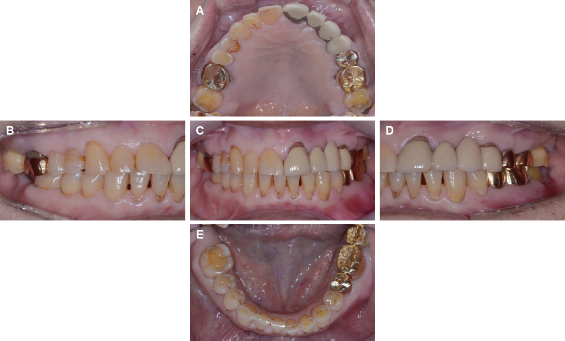
Fig. 1 Intra-oral status in the initial examination. Generalized toothwear was shown. Dentoalveolar abscess was observed around #36. #47 was missing. (A) Maxillary occlusal view, (B) Right lateral view, (C) Frontal view at maximum inter-cuspal position, (D) Left lateral view, (E) Mandibular occlusal view.
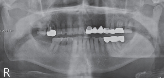
Fig. 2 Initial panoramic radiographic image. Alveolar bone resorption was observed around #16, #21, #25. Distal caries was observed at #17. Periapical radiolucency was observed at #36.

Fig. 3 Initial TMJ series. No evidence of pathologic change. (A) Right side in closed state, (B) Right side in opened state, (C) Left side in closed state, (D) Left side in opened state.

Fig. 4 Vertical dimension analysis. In the Willis analysis, the ratio of the distance from the pupil of the eyes to the parting line of the lips and the distance from the bony shelf under the nose to the bottom of the mandible was 1:0.97. No decrease in the occlusal vertical dimension was observed. (A) Frontal view, (B) Lateral view.
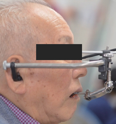
Fig. 5 Face-bow transfer (Right side view).

Fig. 6 Diagnostic wax up without any change in the occlusal vertical dimension. There was enough space for the prosthesis. The distance between both bottoms of labial vestibules of upper and lower casts was 35.0 mm which is the average distance in Korean adults with natural dentition. (A) Right side view, (B) Frontal view, (C) Left side view.
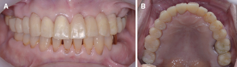
Fig. 7 Provisional restoration after 3 months. Provisional restoration functioned in the patient’s mouth without any problems in mastication, pronunciation, and esthetics. (A) Frontal view, (B) Maxillary occlusal view.
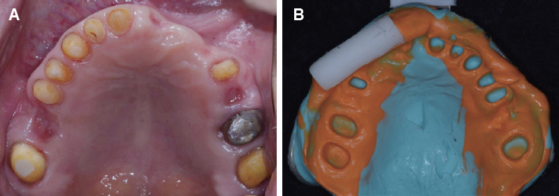
Fig. 8 Final abutment preparation and impression taking for definitive prosthesis. (A) Maxillary abutment preparation, (B) Maxillary abutment impression.
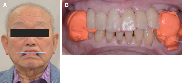
Fig. 9 Registration of inter-occlusal relationship with provisional restoration. (A) Anterior bite registration with posterior portion of provisional restoration displaying the interpupillary line and midline, (B) Posterior bite registration with anterior zig.
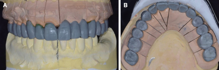
Fig. 10 Full contour wax up for definitive prosthesis. (A) Frontal view, (B) Maxillary occlusal view.
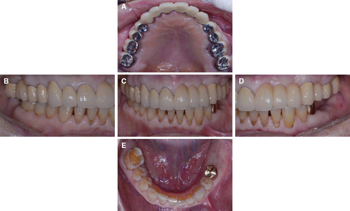
Fig. 11 Definitive prosthesis was delivered. Esthetics and function were restored with the PFM prosthesis. (A) Maxillary occlusal view, (B) Right lateral view, (C) Frontal view at maximum inter-cuspal position, (D) Left lateral view, (E) Mandibular occlusal view.

Fig. 12 Panoramic radiographic image after definitive prosthesis delivery. No additional pathologic change was seen.

Fig. 13 Extra-oral photos after definitive prosthesis delivery. Stable occlusion was observed. (A) Frontal view, (B) Lateral view.
Reference
-
References
1. Donachie MA, Walls AW. Assessment of tooth wear in an ageing population. J Dent. 1995; 23:15764. DOI: 10.1016/0300-5712(95)93573-K.2. Dawson PE. Functional occlusion:from TMJ to smile design. St. Louis: Mosby;2007. p. 430–52.3. Ramfjord SP, Blankenship JR. Increased occlusal vertical dimension in adult monkeys. J Prosthet Dent. 1981; 45:74–83. DOI: 10.1016/0022-3913(81)90015-9.4. Turner KA, Missirlian DM. Restoration of the extremely worn dentition. J Prosthet Dent. 1984; 52:467–74. DOI: 10.1016/0022-3913(84)90326-3.5. Lloyd PM. Fixed prosthodontics and esthetic considerations for the elder adult. J Prosthet Dent. 1994; 72:525–31. DOI: 10.1016/0022-3913(94)90126-0.6. Willis FM. Features of the face involved in full denture prosthesis. Dent Cosmos. 1935; 77:851–4.7. Park JH, Jeong CM, Jeon YC, Lim JS. A study on the occlusal plane and the vertical dimension in Korean adults with natural dentition. J Korean Acad Prosthodont. 2005; 43:41–51.8. McAdam DB. Tooth loading and cuspal guidance in canine and group-function occlusions. J Prosthet Dent. 1976; 35:283–90. DOI: 10.1016/0022-3913(76)90252-3.9. Luke DA, Lucas PW. The significance of cusps. J Oral Rehabil. 1983; 10:197–206. DOI: 10.1111/j.1365-2842.1983.tb00113.x.10. Ingle JI. Alveolar osteoporosis and pulpal death associated with compulsive bruxism. Oral Surg Oral Med Oral Pathol. 1960; 13:1371–81. DOI: 10.1016/0030-4220(60)90302-9.11. Jepson N, Allen F, Moynihan P, Kelly P, Thomason M. Patient satisfaction following restoration of shortened mandibular dental arches in a randomized controlled trial. Int J Prosthodont. 2003; 16:40914.12. Mojon P, Rentsch A, Budtz-Jørgensen E. Relationship between prosthodontics status, caries, and periodontal disease in a geriatric population. Int J Prosthodont. 1995; 8:564–71.13. Budtz-Jørgensen E, Isidor F. A 5-year longitudinal study of cantilevered fixed partial dentures compared with removable partial dentures in a geriatric population. J Prosthet Dent. 1990; 64:42–7. DOI: 10.1016/0022-3913(90)90151-2.14. Heschl A, Haas M, Haas J, Payer M, Wegscheider W, Polansky R. Maxillary rehabilitation of periodontally compromised patients with extensive onepiece fixed prostheses supported by natural teeth:a retrospective longitudinal study. Clin Oral Investig. 2013; 17:45–53. DOI: 10.1007/s00784-012-0681-9. PMID: 22290063.15. Gordon SR, Lloyd PM. Fixed prosthodontics in the elderly population. Life expectancy of fixed restorations, failures, and retreatment methods. Dent Clin North Am. 1992; 36:783–95.16. Shillingburg HT, Sather DA, Wilson EL, Cain JR, Mitchell DL, Blanco LJ, Kessler JC. Fundamentals of fixed prosthodontics. 4th ed. Chicago: Quintes-sence Publishing Co;2012. p. 81–98.17. Landry KE, Johnson PF, Parks VJ, Pelleu GB Jr. A photoelastic study to determine the location of the nonrigid connector in a five-unit intermediate abutment prosthesis. J Prosethet Dent. 1987; 57:454–7. DOI: 10.1016/0022-3913(87)90014-X.18. Reynolds JM. Abutment selection for fixed prosthodontics. J Prosthet Dent. 1968; 19:483–8. DOI: 10.1016/0022-3913(68)90064-4.19. Hung SH, Hung KS, Eick JD, Chappell RP. Marginal fit of porcelain-fused-to-metal and two types of ceramic crown. J Prosthet Dent. 1990; 63:26–31. DOI: 10.1016/0022-3913(90)90260-J.20. Heschl A, Haas M, Haas J, Payer M, Wegscheider W, Polansky R. Maxillary rehabilitation of periodontally compromised patients with extensive onepiece fixed prosthesis supported by natural teeth:a retrospective longitudinal study. Clin Oral Investig. 2013; 17:45–53. DOI: 10.1007/s00784-012-0681-9. PMID: 22290063 .
- Full Text Links
-
- Actions
-
Cited
- CITED
-
- Close
- Share
-
- Similar articles
-
- Implant-supported maxillary full-arch fixed prosthesis opposing mandibular natural dentition: A clinical report
- Using implants for worn dentition with the altered vertical dimension of occlusion based on shortened dental arch concept: a case report
- Full mouth rehabilitation with extremely worn dentition
- Full mouth rehabilitation of the patient with severely worn dentition and limited vertical dimension
- Full-mouth rehabilitation without changing the vertical dimension in patient with worn dentition
