Restor Dent Endod.
2017 Aug;42(3):232-239. 10.5395/rde.2017.42.3.232.
Light-emitting diode assessment of dentinal defects: the role of presumed extraction forces
- Affiliations
-
- 1Department of Endodontics, Universidade Paulista School of Dentistry, Sorocaba, SP, Brazil. coelho_marcelo@yahoo.com.br
- 2Department of Endodontics, University of North Carolina School of Dentistry, Chapel Hill, NC, USA.
- KMID: 2386777
- DOI: http://doi.org/10.5395/rde.2017.42.3.232
Abstract
OBJECTIVES
The evaluation of iatrogenic dentinal defects in extracted teeth may be influenced by extraction forces and prolonged dry times. The purpose of this study was to compare the presence of dentinal defects in freshly extracted, periodontally compromised teeth with those in a group of teeth with uncontrolled extraction forces and storage time.
MATERIALS AND METHODS
The experimental group consisted of eighteen roots obtained from teeth extracted due to periodontal reasons with class II or III mobility. They were kept in saline and sectioned within 1 hour following extraction. The control group consisted of matched root types obtained from an anonymous tooth collection, consistent with previous dentinal defect studies. The slices were obtained at 3, 6, and 9 mm from the apex. The imaging process exposed all specimens to no more than 60 seconds of dry time. The × 12.8 magnification was used for the 9 mm slices and × 19.2 magnification for the 3 mm and 6 mm slices under light-emitting diode (LED) transillumination. The root canal spaces and periodontal tissues were masked to minimize extraneous factors that might influence the evaluators. Chi-square test was used for statistical analysis.
RESULTS
Dentinal defects were detected in 17% of the experimental group teeth, compared to 61% of control teeth (p = 0.015).
CONCLUSIONS
LED transillumination assessment of freshly extracted roots with class II or III mobility showed smaller number of dentinal defects than roots with uncontrolled storage time and extraction forces. The use of freshly extracted roots with mobility should be considered for future dental defect assessment studies.
MeSH Terms
Figure
-
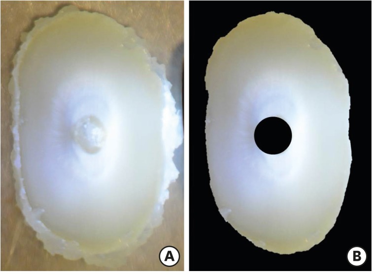
Figure 1 Specimen of group 1 presenting (A) remnants of periodontal ligament and (B) specimen masked.
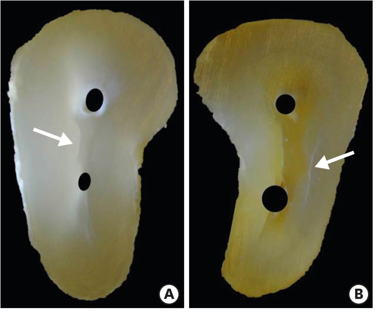
Figure 2 Specimens of (A) group 1 and (B) group 2 presenting dentinal defects (arrows).
Reference
-
1. Bier CA, Shemesh H, Tanomaru-Filho M, Wesselink PR, Wu MK. The ability of different nickel-titanium rotary instruments to induce dentinal damage during canal preparation. J Endod. 2009; 35:236–238. PMID: 19166781.
Article2. Tawil PZ, Saraiya VM, Galicia JC, Duggan DJ. Periapical microsurgery: the effect of root dentinal defects on short- and long-term outcome. J Endod. 2015; 41:22–27. PMID: 25282374.
Article3. Karataş E, Gündüz HA, Kırıcı DO, Arslan H, Topçu MC, Yeter KY. Dentinal crack formation during root canal preparations by the twisted file adaptive, ProTaper Next, ProTaper Universal, and WaveOne instruments. J Endod. 2015; 41:261–264. PMID: 25476974.
Article4. Shemesh H, Wesselink PR, Wu MK. Incidence of dentinal defects after root canal filling procedures. Int Endod J. 2010; 43:995–1000. PMID: 20722755.
Article5. Topçuoğlu HS, Demirbuga S, Tuncay Ö, Pala K, Arslan H, Karataş E. The effects of Mtwo, R-Endo, and D-RaCe retreatment instruments on the incidence of dentinal defects during the removal of root canal filling material. J Endod. 2014; 40:266–270. PMID: 24461416.
Article6. Shemesh H, Bier CA, Wu MK, Tanomaru-Filho M, Wesselink PR. The effects of canal preparation and filling on the incidence of dentinal defects. Int Endod J. 2009; 42:208–213. PMID: 19228210.
Article7. Coelho MS, Card SJ, Tawil PZ. Visualization enhancement of dentinal defects by using light-emitting diode transillumination. J Endod. 2016; 42:1110–1113. PMID: 27178248.
Article8. Rose E, Svec T. An evaluation of apical cracks in teeth undergoing orthograde root canal instrumentation. J Endod. 2015; 41:2021–2024. PMID: 26472677.
Article9. Arias A, Lee YH, Peters CI, Gluskin AH, Peters OA. Comparison of 2 canal preparation techniques in the induction of microcracks: a pilot study with cadaver mandibles. J Endod. 2014; 40:982–985. PMID: 24935548.
Article10. Coelho MS, Card SJ, Tawil PZ. Light-emitting diode assessment of dentinal defects after root canal preparation with Profile, TRUShape, and WaveOne Gold Systems. J Endod. 2016; 42:1393–1396. PMID: 27421973.
Article11. De-Deus G, Silva EJ, Marins J, Souza E, Neves AA, Gonçalves Belladonna F, Alves H, Lopes RT, Versiani MA. Lack of causal relationship between dentinal microcracks and root canal preparation with reciprocation systems. J Endod. 2014; 40:1447–1450. PMID: 25146030.
Article12. Versiani MA, Souza E, De-Deus G. Critical appraisal of studies on dentinal radicular microcracks in endodontics: methodological issues, contemporary concepts, and future perspectives. Endod Topics. 2015; 33:87–156.13. Miller PD Jr. A classification of marginal tissue recession. Int J Periodontics Restorative Dent. 1985; 5:8–13.14. Saber SE, Schäfer E. Incidence of dentinal defects after preparation of severely curved root canals using the Reciproc single-file system with and without prior creation of a glide path. Int Endod J. 2016; 49:1057–1064. PMID: 26426069.
Article15. Abou El Nasr HM, Abd El Kader KG. Dentinal damage and fracture resistance of oval roots prepared with single-file systems using different kinematics. J Endod. 2014; 40:849–851. PMID: 24862715.
Article16. Bürklein S, Tsotsis P, Schäfer E. Incidence of dentinal defects after root canal preparation: reciprocating versus rotary instrumentation. J Endod. 2013; 39:501–504. PMID: 23522545.
Article17. Arslan H, Karataş E, Çapar ID, Ozsu D, Doğanay E. Effect of ProTaper Universal, Endoflare, Revo-S, HyFlex coronal flaring instruments, and Gates Glidden drills on crack formation. J Endod. 2014; 40:1681–1683. PMID: 25127932.18. Adorno CG, Yoshioka T, Suda H. The effect of root preparation technique and instrumentation length on the development of apical root cracks. J Endod. 2009; 35:389–392. PMID: 19249601.
Article19. Jamleh A, Komabayashi T, Ebihara A, Nassar M, Watanabe S, Yoshioka T, Miyara K, Suda H. Root surface strain during canal shaping and its influence on apical microcrack development: a preliminary investigation. Int Endod J. 2015; 48:1103–1111. PMID: 25377258.
Article20. Ceyhanli KT, Erdilek N, Tatar I, Celik D. Comparison of ProTaper, RaCe and Safesider instruments in the induction of dentinal microcracks: a micro-CT study. Int Endod J. 2016; 49:684–689. PMID: 26172031.
Article21. Chan CP, Tseng SC, Lin CP, Huang CC, Tsai TP, Chen CC. Vertical root fracture in nonendodontically treated teeth--a clinical report of 64 cases in Chinese patients. J Endod. 1998; 24:678–681. PMID: 10023252.22. Tsesis I, Rosen E, Tamse A, Taschieri S, Kfir A. Diagnosis of vertical root fractures in endodontically treated teeth based on clinical and radiographic indices: a systematic review. J Endod. 2010; 36:1455–1458. PMID: 20728708.
Article23. Lim H, Li FC, Friedman S, Kishen A. Residual microstrain in root dentin after canal instrumentation measured with digital moiré interferometry. J Endod. 2016; 42:1397–1402. PMID: 27430943.
Article24. Hin ES, Wu MK, Wesselink PR, Shemesh H. Effects of self-adjusting file, Mtwo, and ProTaper on the root canal wall. J Endod. 2013; 39:262–264. PMID: 23321242.
Article25. Kansal R, Rajput A, Talwar S, Roongta R, Verma M. Assessment of dentinal damage during canal preparation using reciprocating and rotary files. J Endod. 2014; 40:1443–1446. PMID: 25146029.
Article26. Çapar ID, Uysal B, Ok E, Arslan H. Effect of the size of the apical enlargement with rotary instruments, single-cone filling, post space preparation with drills, fiber post removal, and root canal filling removal on apical crack initiation and propagation. J Endod. 2015; 41:253–256. PMID: 25433969.27. Adorno CG, Yoshioka T, Jindan P, Kobayashi C, Suda H. The effect of endodontic procedures on apical crack initiation and propagation ex vivo . Int Endod J. 2013; 46:763–768. PMID: 23402216.28. Pedullà E, Genovesi F, Rapisarda S, La Rosa GR, Grande NM, Plotino G, Adorno CG. Effects of 6 single-file systems on dentinal crack formation. J Endod. 2017; 43:456–461. PMID: 28131416.
Article29. Ivancik J, Majd H, Bajaj D, Romberg E, Arola D. Contributions of aging to the fatigue crack growth resistance of human dentin. Acta Biomater. 2012; 8:2737–2746. PMID: 22484693.
Article30. Mireku AS, Romberg E, Fouad AF, Arola D. Vertical fracture of root filled teeth restored with posts: the effects of patient age and dentine thickness. Int Endod J. 2010; 43:218–225. PMID: 20158533.
Article31. PradeepKumar AR. Shemesh H, Jothilatha S, Vijayabharathi R, Jayalakshmi S, Kishen A. Diagnosis of vertical root fractures in restored endodontically treated teeth: a time-dependent retrospective cohort study. J Endod. 2016; 42:1175–1180. PMID: 27339633.
- Full Text Links
-
- Actions
-
Cited
- CITED
-
- Close
- Share
-
- Similar articles
-
- The shear bond strength and adhesive failure pattern in bracket bonding with different light-curing methods
- Topical Photodynamic Therapy for Treatment of Actinic Keratosis Using Light-Emitting Diode (LED) Device
- Transfer printing based manufacturing process for flexible micro-light emitting diode phototherapy devices
- Evaluation of High-power Light Emitting Diode Curing Light on Sealant Polymerization
- The Effect of 650 nm Red Light Phototherapy on Acne Vulgaris
