MR Imaging for Staging of Cervical Carcinoma: Update
- Affiliations
-
- 1Department of Radiology, Dong-A University College of Medicine, Dong-A University Hospital, Busan, Korea. cerub@naver.com
- KMID: 2386742
- DOI: http://doi.org/10.3348/jksr.2017.77.2.67
Abstract
- Uterine cervical cancer is globally the third most common cancer among women, and shows high mortality with invasive cervical carcinoma. Early detection of the disease, its correct staging, and treatment are therefore of great importance. The staging system updated in 2009 by the International Federation of Gynecology and Obstetrics (FIGO), is commonly used for planning the treatment. However, there are significant inaccuracies in the FIGO staging system. Accurate tumor staging is very important to decide the treatment strategy. Although not included in the staging system, magnetic resonance (MR) imaging is a valuable tool for local staging of the disease, and is useful in assessing the spread of the tumor and metastatic lymph nodes, thereby becoming a more accurate substitute for clinical staging of cervical carcinoma. In addition, it is capable of assessing the disease response to surgery or chemoradiation. This review briefly describes the role of MR imaging and the basic MR scanning protocol in evaluating cervical carcinoma. The MR findings with staging, and MR evaluation of treatment response, are further addressed.
MeSH Terms
Figure
-
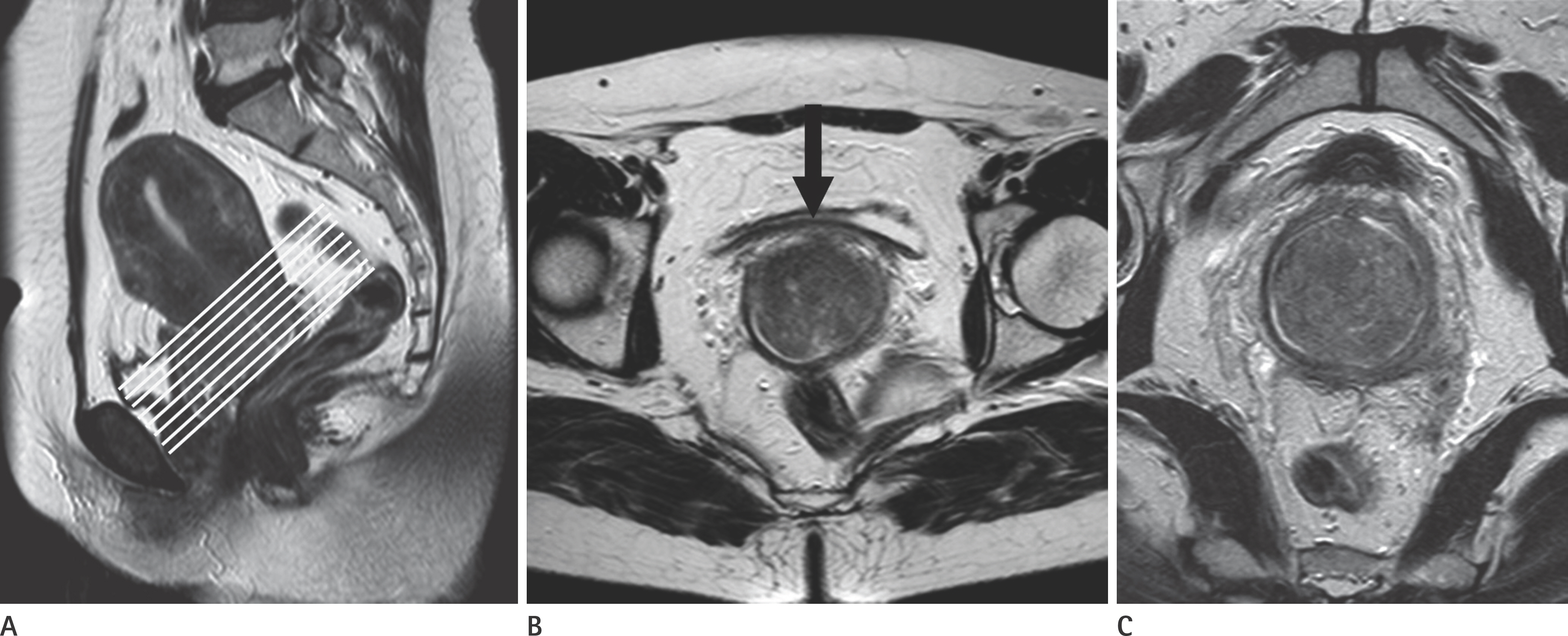
Fig. 1. Axial oblique T2-weighted MR image. A. Sagittal T2-weighted MR image shows a heterogeneous, intermediate signal intensity mass in the uterine cervix. Lines indicate scan direction of oblique axial images, which are obtained perpendicular to the long axis of cervix. B. Axial T2-weighted image shows that parametrial invasion is not clear in the anterior aspect of the cervix (arrow). C. Oblique axial T2-weighted image clearly excludes the extension to the parametria, which provides more accurate assessment of stromal and parametrial invasion, as compared with T2-weighted image.

Fig. 2. Stage IB1 cervical carcinoma in a 59-year-old woman. A, B. Sagittal (A) and axial (B) T2-weighted MR images show no abnormal signal intensity lesions in the uterine cervix. C. Axial contrast-enhanced T1-weighted MR image shows a small enhancing tumor (arrow) in the cervix. D. A photograph of gross specimen of the uterine cervix reveals a small nodular mass (arrow) within the endocervix, which is staged as IB1 disease after radical hysterectomy.
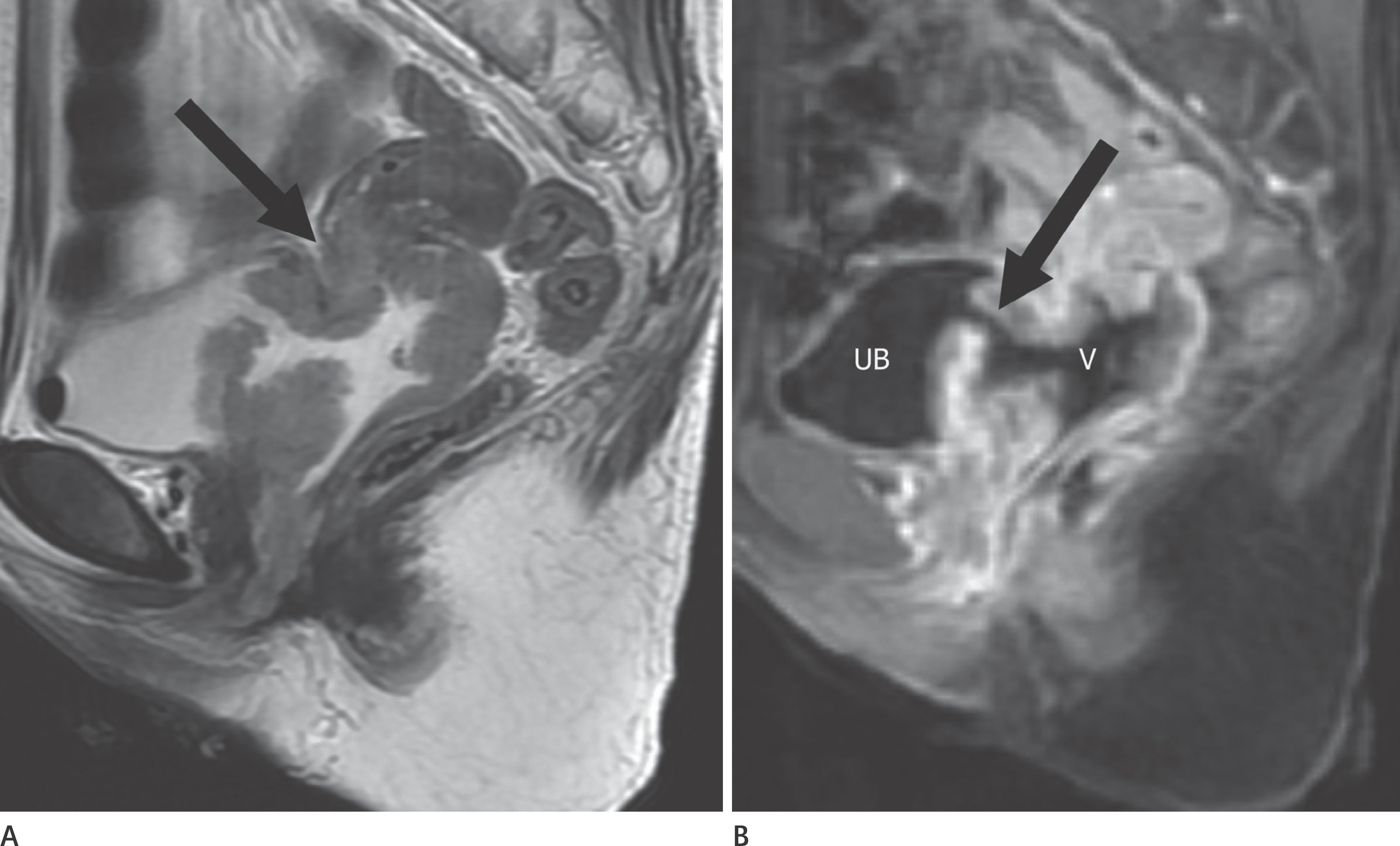
Fig. 3. Stage IVA cervical carcinoma in a 58-year-old woman. A. Sagittal T2-weighted MR image shows a bulky tumor (arrow) with invasion to the posterior wall of the bladder. B. Sagittal contrast-enhanced T1-weighted MR image clearly demonstrates an enhancing mass with fistulous tract (arrow) between the urinary bladder (UB) and vagina (V), a finding indicative of vesicovaginal fistula.
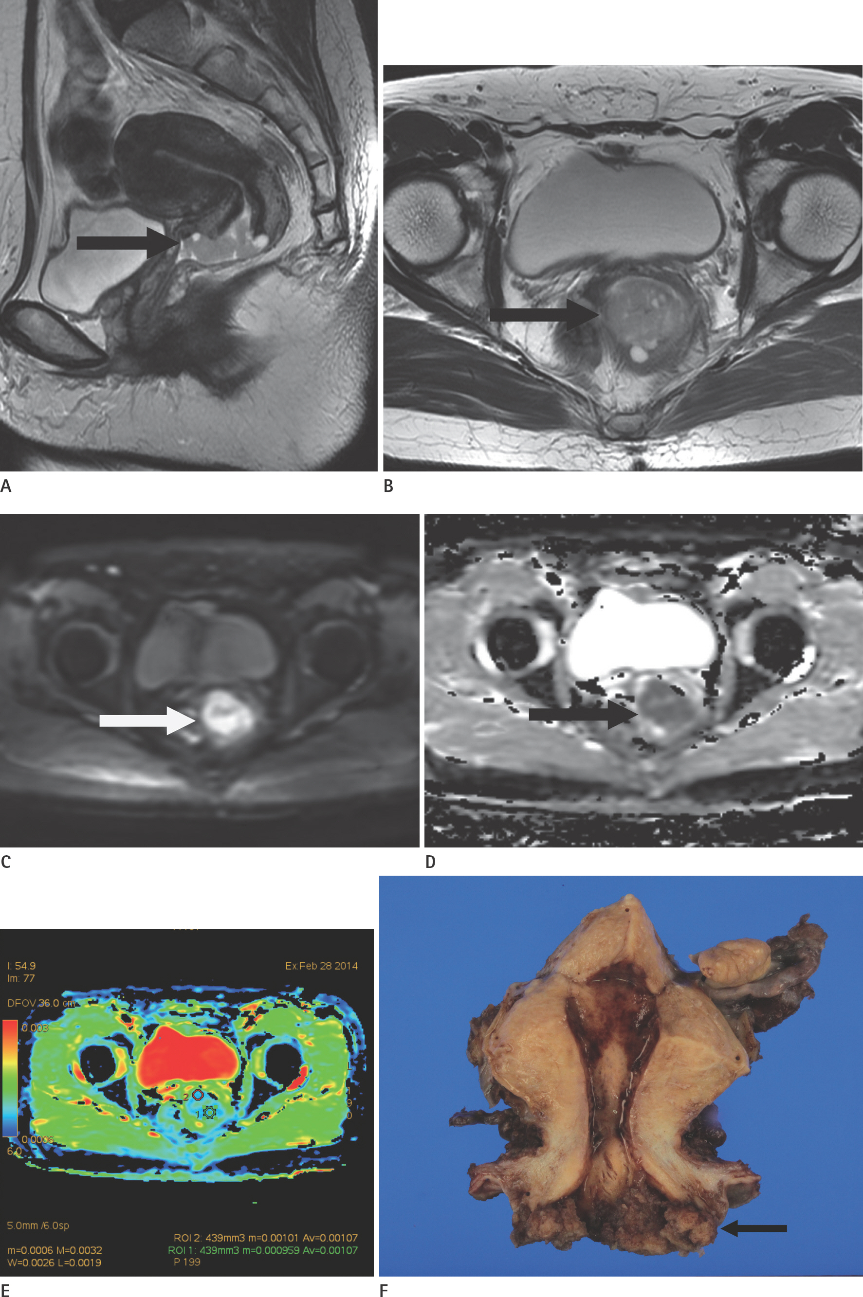
Fig. 4. Stage IB1 cervical carcinoma in a 51-year-old woman. A, B. Sagittal (A) and axial oblique (B) T2-weighted MR images show an exophytic hyperintense mass (arrows) arising from external os of the uterine cervix, without parametrial invasion. Stage IB1 cervical carcinoma in a 51-year-old woman. C-E. Axial DWI (C) and ADC (D, E) maps show a hyperintense mass (white arrow) with diffusion restriction (black arrow) and decreased mean ADC value (1.070 × 10⁻³ mm2/s). F. A photograph of the gross specimen of the uterine cervix demonstrates a polypoid mass with exophytic growing from external os of the uter-ine cervix (arrow). ADC = apparent diffusion coefficient, DWI = diffusion-weighted imaging

Fig. 5. Stage IIA2 cervical carcinoma in a 63-year-old woman. A. Sagittal T2-weighted MR image shows a hyperintense exophytic growing mass with extension to the upper half of the anterior vaginal wall (arrow). B. A photograph of the gross specimen of the uterine cervix reveals an ill-defined, infiltrative mass (4.5 × 2 cm) with extension to the upper anteri-or vagina (arrow), after radical hysterectomy.
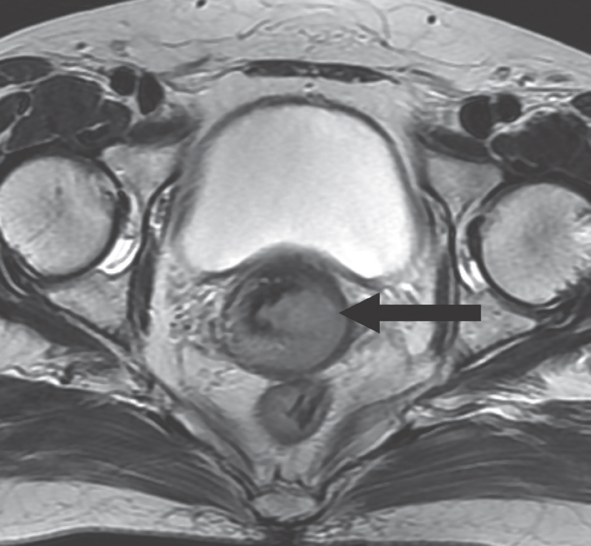
Fig. 6. Stage IIB cervical carcinoma in a 63-year-old woman. Axial T2-weighted MR image shows an intermediate signal intensity mass with focal disruption of hypointense stromal ring in the left side (arrow), a finding indicative of stromal invasion.
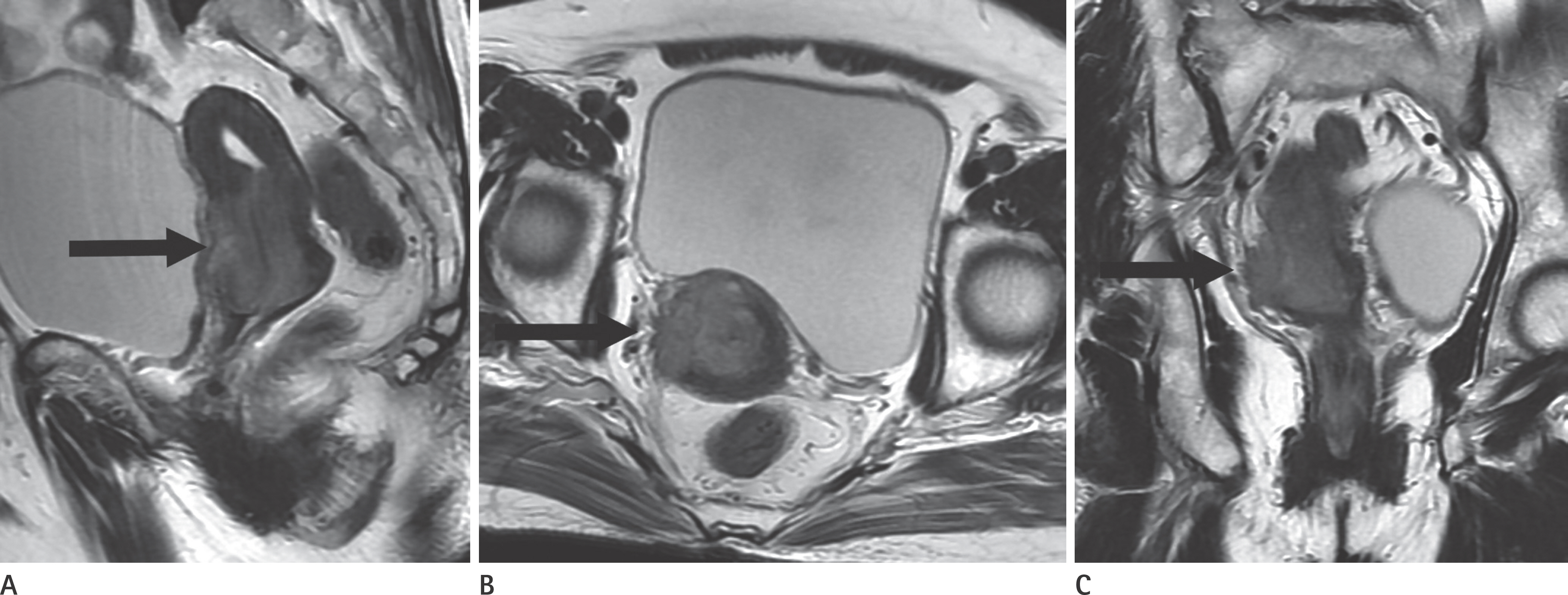
Fig. 7. Stage IIB cervical carcinoma in a 77-year-old woman. A. Sagittal T2-weighted MR image shows a relatively ill-defined, hyperintense mass (arrow) in the cervix. B, C. Axial oblique (B) T2-weighted MR image shows disruption of hypointense ring and spiculated tumor-parametrial interface, a finding con-sistent with parametrial invasion (arrow). This finding is also clearly seen on coronal (C) T2-weighted MR image (arrow).
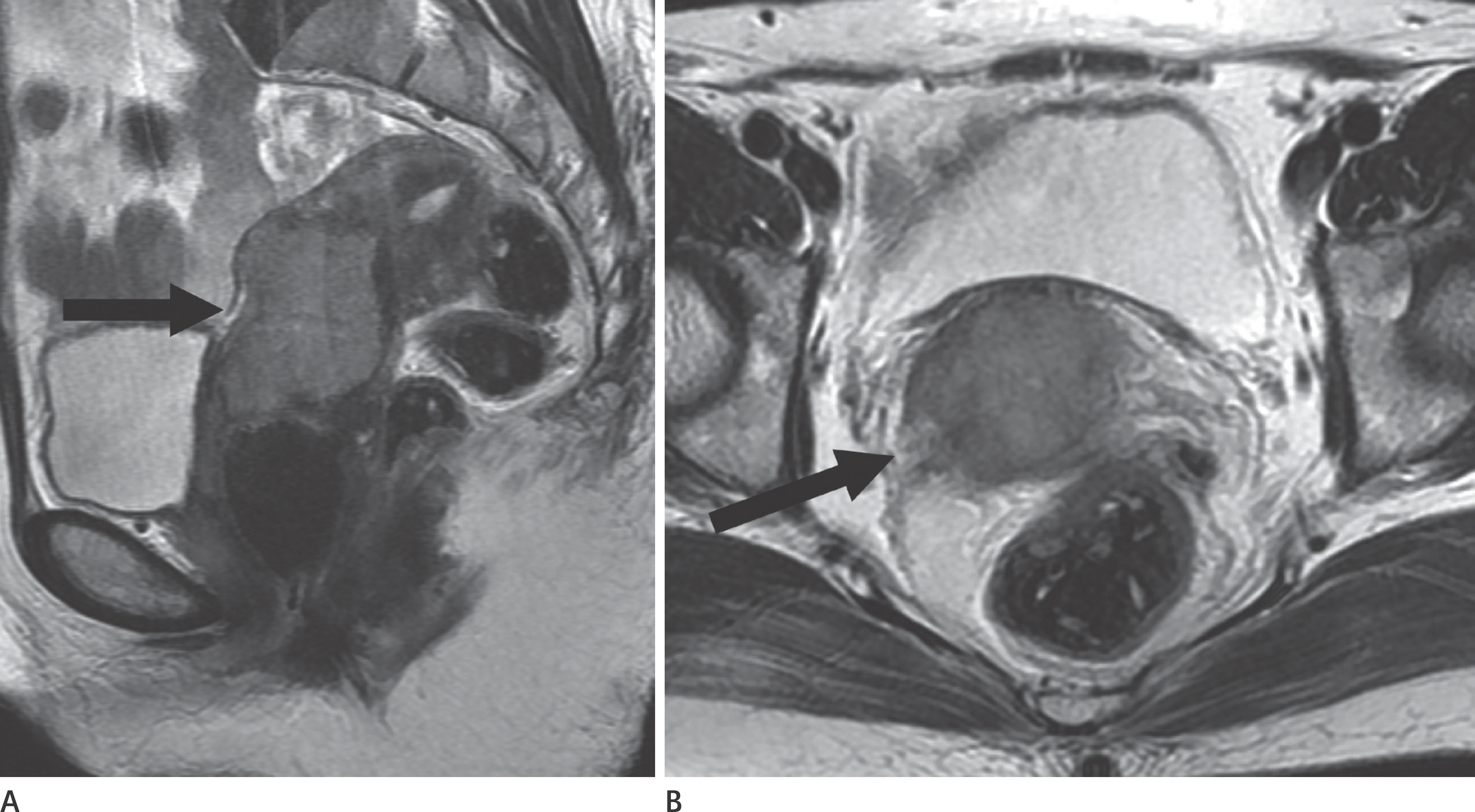
Fig. 8. Stage IB2 cervical carcinoma in a 46-year-old woman. A. Sagittal T2-weighted MR image shows a lobulated hyperintense mass (arrow) in the endocervix. B. Axial T2-weighted MR image shows irregular and spiculated tumor-parametrial interface (arrow), suggestive of clinical stage IIB disease. How-ever, this lesion was overstaged and proven stage IB2 after radical hysterectomy.
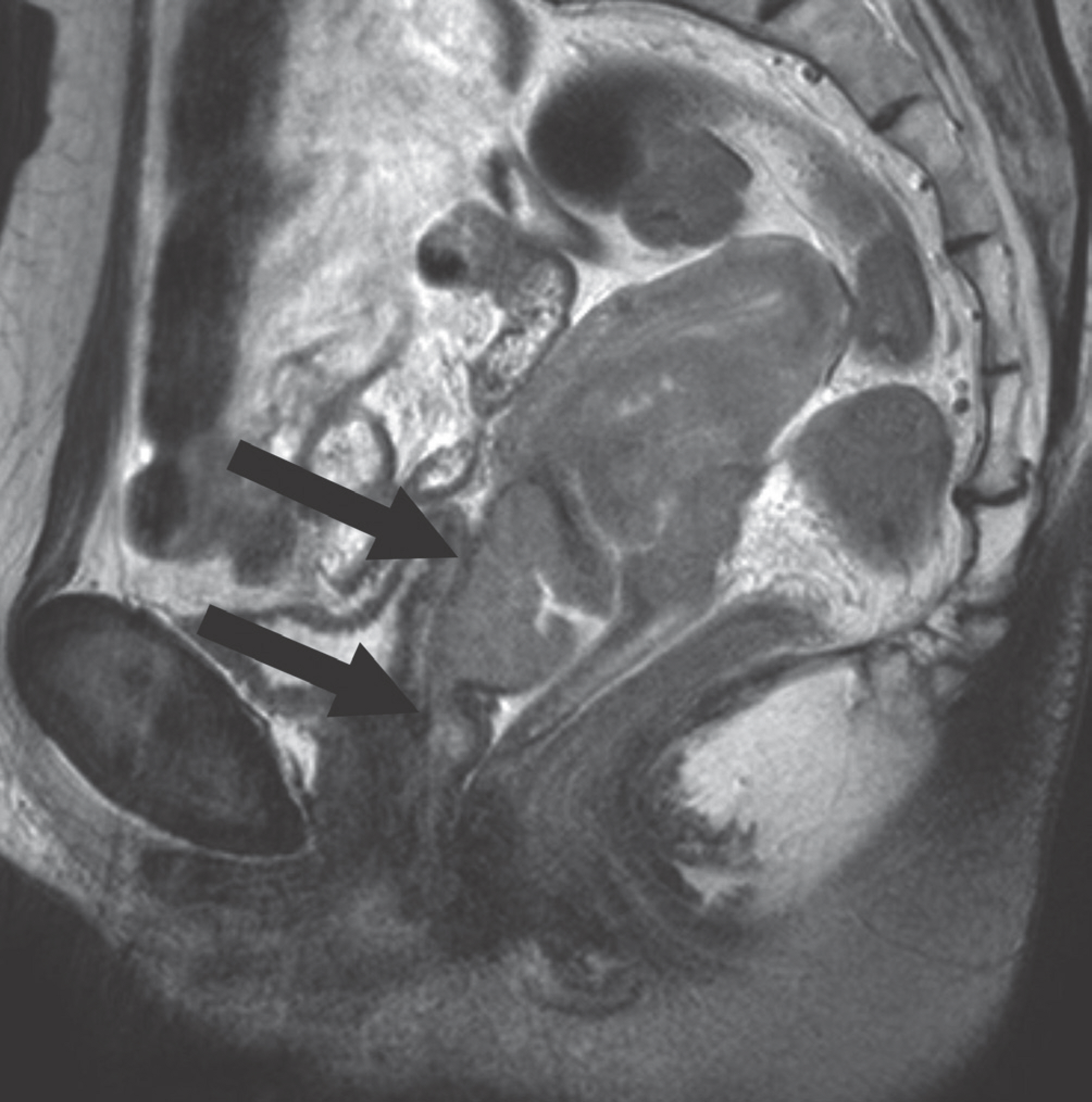
Fig. 9. Stage IIIA cervical carcinoma in a 64-year-old woman. Sagittal T2-weighted MR image shows a hyperintense mass and extension to anterior upper and lower vaginal wall (arrows), which is a favorable finding of vaginal invasion. However, hypointense posterior wall of the blad-der is intact.

Fig. 10. Stage IIIB cervical carcinoma in a 75-year-old woman. A, B. Axial (A) and coronal (B) T2-weighted MR images show intermediate signal intensity mass in the cervix, extending into the right lower ure-ter (arrows). C. Axial T2-weighted MR image obtained at the renal hilar level shows hydronephrosis (arrow) in the right kidney.

Fig. 11. Stage IVA cervical carcinoma in a 48-year-old woman. Sagittal T2-weighted MR image shows tumor extension into the posterior wall of the bladder and anterior wall of the rectosigmoid colon, findings con-sistent with bladder and rectal invasion (arrows).
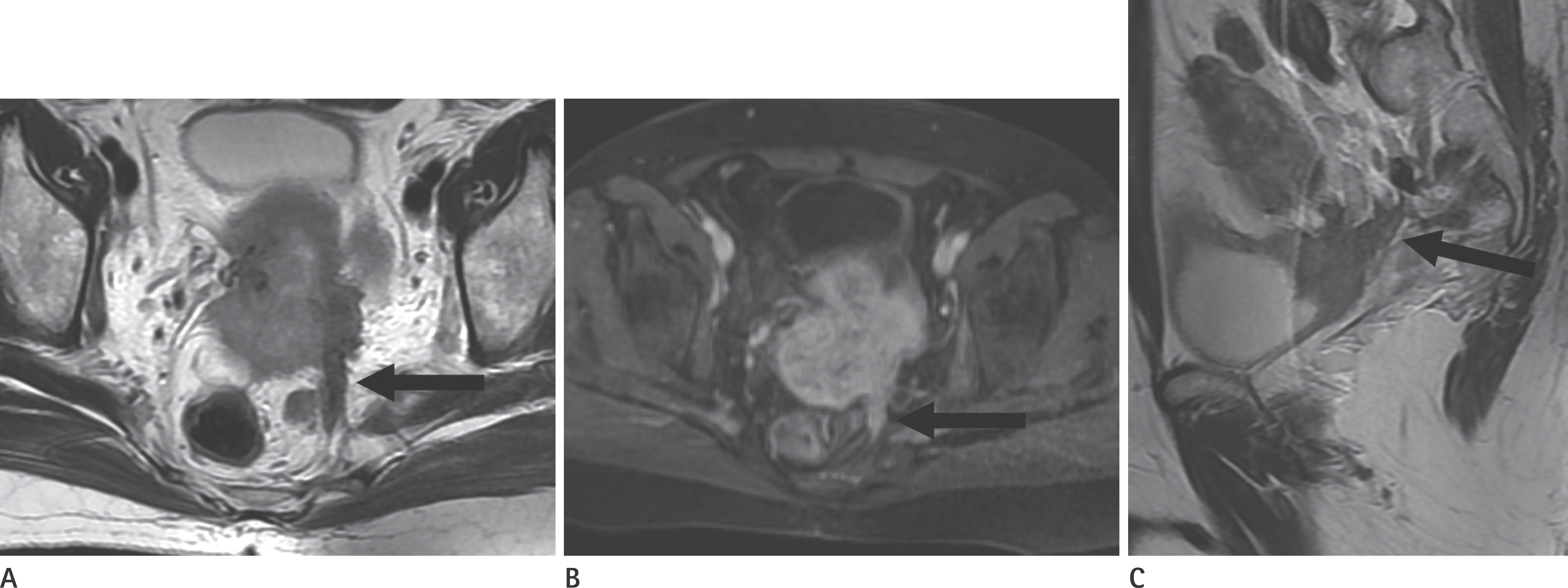
Fig. 12. Stage IVA cervical carcinoma in a 52-year-old woman. A, B. Axial T2-weighted (A) and contrast-enhanced T1-weighted (B) MR images show an intermediate signal intensity mass with extension to the uterosacral ligament (arrow) posteriorly. C. Coronal T2-weighted MR image shows tumor invasion to the posterior wall of the bladder as well as the uterosacral ligament (arrow).
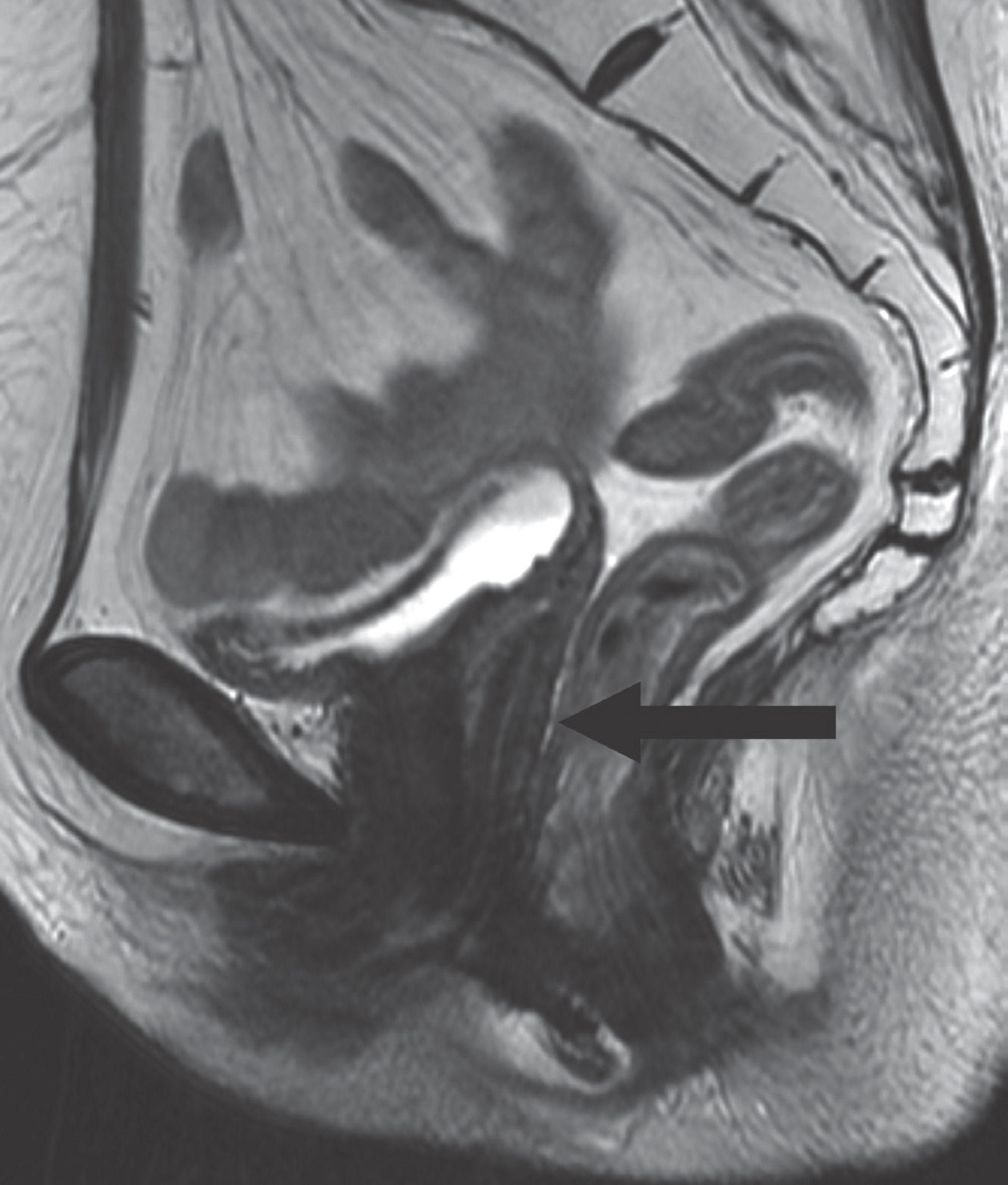
Fig. 13. Total hysterectomy for stage IA1 cervical carcinoma in a 73-year-old woman. Sagittal T2-weighted MR image shows absence of the uter-us with no abnormal signal intensity mass in the vaginal cuff and nor-mal appearing smooth, low signal intensity vaginal wall (arrow).
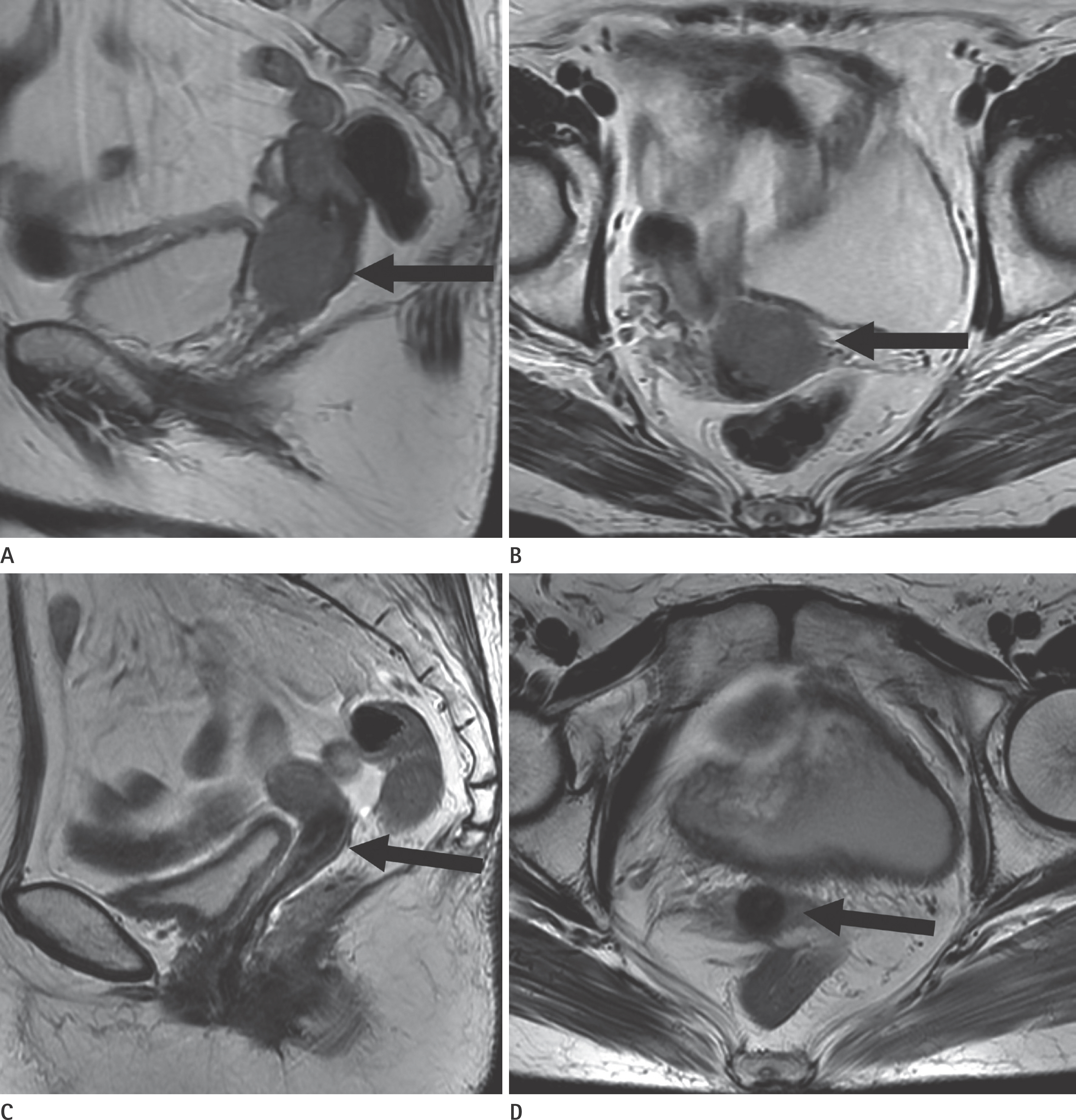
Fig. 14. Complete response after radiation treatment for stage IIB cervical carcinoma in a 67-year-old woman. A, B. Pre-treatment sagittal (A) and axial (B) T2-wighted images show an intermediate signal intensity mass (arrows) with invasion to the upper vagina and parametrium, a finding indicative of stage IIB disease. C, D. Following radiation treatment, sagittal (C) and axial (D) T2-wighted images show diffuse, homogeneous low signal intensity cervical stro-ma, and reconstitution of the normal zonal anatomy in the uterine cervix (arrows), a finding consistent with complete response.

Fig. 15. A 42-year-old woman with a recurrent cervical cancer after surgical treatment. A, B. Following radical hysterectomy, sagittal (A) and axial (B) T2-weighted MR images show intermediate signal intensity mass in the vaginal cuff (arrows). C. Post-contrast T1-weighted MR image shows good enhancement of the mass in the vaginal cuff (white arrow), a finding consistent with a re-current cancer. D, E. Axial DWI (D) and ADC (E) maps show a hyperintense mass (white arrow) with diffusion restriction (black arrow) in the corresponding area. ADC = apparent diffusion coefficient, DWI = diffusion-weighted imaging
Reference
-
1.Akin O., Mironov S., Pandit-Taskar N., Hann LE. Imaging of uterine cancer. Radiol Clin North Am. 2007. 45:167–182.
Article2.Kaur H., Silverman PM., Iyer RB., Verschraegen CF., Eifel PJ., Charnsangavej C. Diagnosis, staging, and surveillance of cervi-cal carcinoma. AJR Am J Roentgenol. 2003. 180:1621–1631.
Article3.Okamoto Y., Tanaka YO., Nishida M., Tsunoda H., Yoshikawa H., Itai Y. MR imaging of the uterine cervix: imaging-patholog-ic correlation. Radiographics. 2003. 23:425–445. quiz 534-535.
Article4.Heller PB., Maletano JH., Bundy BN., Barnhill DR., Okagaki T. Clinical-pathologic study of stage IIB, III, and IVA carcinoma of the cervix: extended diagnostic evaluation for paraaortic node metastasis--a Gynecologic Oncology Group Study. Gy-necol Oncol. 1990. 38:425–430.
Article5.Delgado G., Bundy BN., Fowler WC Jr., Stehman FB., Sevin B., Creasman WT, et al. A prospective surgical pathological study of stage I squamous carcinoma of the cervix: a Gynecologic Oncology Group Study. Gynecol Oncol. 1989. 35:314–320.
Article6.Heaps JM., Berek JS. Surgical staging of cervical cancer. Clin Obstet Gynecol. 1990. 33:852–862.
Article7.Pecorelli S. Revised FIGO staging for carcinoma of the vulva, cervix, and endometrium. Int J Gynaecol Obstet. 2009. 105:103–104.
Article8.Pecorelli S., Zigliani L., Odicino F. Revised FIGO staging for carcinoma of the cervix. Int J Gynaecol Obstet. 2009. 105:107–108.
Article9.Lagasse LD., Creasman WT., Shingleton HM., Ford JH., Blessing JA. Results and complications of operative staging in cervi-cal cancer: experience of the Gynecologic Oncology Group. Gynecol Oncol. 1980. 9:90–98.
Article10.Subak LL., Hricak H., Powell CB., Azizi L., Stern JL. Cervical car-cinoma: computed tomography and magnetic resonance imaging for preoperative staging. Obstet Gynecol. 1995. 86:43–50.
Article11.Piver MS., Chung WS. Prognostic significance of cervical lesion size and pelvic node metastases in cervical carcinoma. Obstet Gynecol. 1975. 46:507–510.12.Hricak H., Yu KK. Radiology in invasive cervical cancer. AJR Am J Roentgenol. 1996. 167:1101–1108.
Article13.Tirumani SH., Shanbhogue AK., Prasad SR. Current concepts in the diagnosis and management of endometrial and cer-vical carcinomas. Radiol Clin North Am. 2013. 51:1087–1110.
Article14.Vidaurreta J., Bermúdez A., di Paola G., Sardi J. Laparoscopic staging in locally advanced cervical carcinoma: a new pos-sible philosophy? Gynecol Oncol. 1999. 75:366–371.
Article15.LaPolla JP., Schlaerth JB., Gaddis O., Morrow CP. The influence of surgical staging on the evaluation and treatment of patients with cervical carcinoma. Gynecol Oncol. 1986. 24:194–206.
Article16.Zand KR., Reinhold C., Abe H., Maheshwari S., Mohamed A., Upegui D. Magnetic resonance imaging of the cervix. Can-cer Imaging. 2007. 7:69–76.
Article17.Pannu HK., Corl FM., Fishman EK. CT evaluation of cervical cancer: spectrum of disease. Radiographics. 2001. 21:1155–1168.
Article18.Bipat S., Glas AS., van der Velden J., Zwinderman AH., Bossuyt PM., Stoker J. Computed tomography and magnetic reso-nance imaging in staging of uterine cervical carcinoma: a systematic review. Gynecol Oncol. 2003. 91:59–66.
Article19.Hricak H., Gatsonis C., Coakley FV., Snyder B., Reinhold C., Schwartz LH, et al. Early invasive cervical cancer: CT and MR imaging in preoperative evaluation-ACRIN/GOG comparative study of diagnostic performance and interobserver vari-ability. Radiology. 2007. 245:491–498.
Article20.Nicolet V., Carignan L., Bourdon F., Prosmanne O. MR imaging of cervical carcinoma: a practical staging approach. Radio-graphics. 2000. 20:1539–1549.
Article21.Balleyguier C., Sala E., Da Cunha T., Bergman A., Brkljacic B., Danza F, et al. Staging of uterine cervical cancer with MRI: guidelines of the European Society of Urogenital Radiology. Eur Radiol. 2011. 21:1102–1110.
Article22.Scheidler J., Heuck AF., Steinborn M., Kimmig R., Reiser MF. Parametrial invasion in cervical carcinoma: evaluation of detection at MR imaging with fat suppression. Radiology. 1998. 206:125–129.
Article23.Freeman SJ., Aly AM., Kataoka MY., Addley HC., Reinhold C., Sala E. The revised FIGO staging system for uterine malignan-cies: implications for MR imaging. Radiographics. 2012. 32:1805–1827.
Article24.Hori M., Kim T., Murakami T., Imaoka I., Onishi H., Tomoda K, et al. Uterine cervical carcinoma: preoperative staging with 3.0-T MR imaging--comparison with 1.5-T MR imaging. Radiol-ogy. 2009. 251:96–104.
Article25.Shin YR., Rha SE., Choi BG., Oh SN., Park MY., Byun JY. Uterine cervical carcinoma: a comparison of two- and three-dimensional T2-weighted turbo spin-echo MR imaging at 3.0 T for image quality and local-regional staging. Eur Radiol. 2013. 23:1150–1157.26.Choi SH., Kim SH., Choi HJ., Park BK., Lee HJ. Preoperative mag-netic resonance imaging staging of uterine cervical carci-noma: results of prospective study. J Comput Assist Tomogr. 2004. 28:620–627.27.Sala E., Wakely S., Senior E., Lomas D. MRI of malignant neo-plasms of the uterine corpus and cervix. AJR Am J Roent-genol. 2007. 188:1577–1587.
Article28.Van Vierzen PB., Massuger LF., Ruys SH., Barentsz JO. Fast dy-namic contrast enhanced MR imaging of cervical carcinoma. Clin Radiol. 1998. 53:183–192.
Article29.Yuh WT., Mayr NA., Jarjoura D., Wu D., Grecula JC., Lo SS, et al. Predicting control of primary tumor and survival by DCE MRI during early therapy in cervical cancer. Invest Radiol. 2009. 44:343–350.
Article30.Zahra MA., Tan LT., Priest AN., Graves MJ., Arends M., Crawford RA, et al. Semiquantitative and quantitative dynamic contrast-enhanced magnetic resonance imaging measurements predict radiation response in cervix cancer. Int J Radiat On-col Biol Phys. 2009. 74:766–773.
Article31.Kim JH., Kim CK., Park BK., Park SY., Huh SJ., Kim B. Dynamic contrast-enhanced 3-T MR imaging in cervical cancer before and after concurrent chemoradiotherapy. Eur Radiol. 2012. 22:2533–2539.
Article32.Nougaret S., Tirumani SH., Addley H., Pandey H., Sala E., Rein-hold C. Pearls and pitfalls in MRI of gynecologic malignan-cy with diffusion-weighted technique. AJR Am J Roentgen-ol. 2013. 200:261–276.
Article33.Hoogendam JP., Klerkx WM., de Kort GA., Bipat S., Zweemer RP., Sie-Go DM, et al. The influence of the b-value combination on apparent diffusion coefficient based differentiation between malignant and benign tissue in cervical cancer. J Magn Reson Imaging. 2010. 32:376–382.
Article34.Chen J., Zhang Y., Liang B., Yang Z. The utility of diffusion-weighted MR imaging in cervical cancer. Eur J Radiol. 2010. 74:e101–e106.
Article35.Liu Y., Bai R., Sun H., Liu H., Zhao X., Li Y. Diffusion-weighted imaging in predicting and monitoring the response of uter-ine cervical cancer to combined chemoradiation. Clin Ra-diol. 2009. 64:1067–1074.
Article36.Payne GS., Schmidt M., Morgan VA., Giles S., Bridges J., Ind T, et al. Evaluation of magnetic resonance diffusion and spec-troscopy measurements as predictive biomarkers in stage 1 cervical cancer. Gynecol Oncol. 2010. 116:246–252.
Article37.Heo SH., Shin SS., Kim JW., Lim HS., Jeong YY., Kang WD, et al. Pre-treatment diffusion-weighted MR imaging for predicting tumor recurrence in uterine cervical cancer treated with concurrent chemoradiation: value of histogram analysis of apparent diffusion coefficients. Korean J Radiol. 2013. 14:616–625.
Article38.Fu ZZ., Peng Y., Cao LY., Chen YS., Li K., Fu BH. Value of appar-ent diffusion coefficient (ADC) in assessing radiotherapy and chemotherapy success in cervical cancer. Magn Reson Imaging. 2015. 33:516–524.
Article39.Kim HS., Kim CK., Park BK., Huh SJ., Kim B. Evaluation of ther-apeutic response to concurrent chemoradiotherapy in patients with cervical cancer using diffusion-weighted MR im-aging. J Magn Reson Imaging. 2013. 37:187–193.
Article40.Kim SH., Choi BI., Lee HP., Kang SB., Choi YM., Han MC, et al. Uterine cervical carcinoma: comparison of CT and MR findings. Radiology. 1990. 175:45–51.
Article41.Togashi K., Nishimura K., Sagoh T., Minami S., Noma S., Fujisawa I, et al. Carcinoma of the cervix: staging with MR imaging. Radiology. 1989. 171:245–251.
Article42.Lien HH., Blomlie V., Kjørstad K., Abeler V., Kaalhus O. Clinical stage I carcinoma of the cervix: value of MR imaging in de-termining degree of invasiveness. AJR Am J Roentgenol. 1991. 156:1191–1194.
Article43.Rockall AG., Ghosh S., Alexander-Sefre F., Babar S., Younis MT., Naz S, et al. Can MRI rule out bladder and rectal invasion in cervical cancer to help select patients for limited EUA? Gy-necol Oncol. 2006. 101:244–249.
Article44.Shiraiwa M., Joja I., Asakawa T., Okuno K., Shibutani O., Aka-matsu N, et al. Cervical carcinoma: efficacy of thin-section oblique axial T2-weighted images for evaluating parametri-al invasion. Abdom Imaging. 1999. 24:514–519.
Article45.Sheu M., Chang C., Wang J., Yen M. MR staging of clinical stage I and IIa cervical carcinoma: a reappraisal of efficacy and pitfalls. Eur J Radiol. 2001. 38:225–231.
Article46.Hricak H., Lacey CG., Sandles LG., Chang YC., Winkler ML., Stern JL. Invasive cervical carcinoma: comparison of MR imaging and surgical findings. Radiology. 1988. 166:623–631.
Article47.Kim SH., Han MC. Invasion of the urinary bladder by uterine cervical carcinoma: evaluation with MR imaging. AJR Am J Roentgenol. 1997. 168:393–397.
Article48.Son H., Kositwattanarerk A., Hayes MP., Chuang L., Rahaman J., Heiba S, et al. PET/CT evaluation of cervical cancer: spec-trum of disease. Radiographics. 2010. 30:1251–1268.
Article49.Sakuragi N., Satoh C., Takeda N., Hareyama H., Takeda M., Ya-mamoto R, et al. Incidence and distribution pattern of pel-vic and paraaortic lymph node metastasis in patients with Stages IB, IIA, and IIB cervical carcinoma treated with radi-cal hysterectomy. Cancer. 1999. 85:1547–1554.
Article50.Kim SH., Kim SC., Choi BI., Han MC. Uterine cervical carcino-ma: evaluation of pelvic lymph node metastasis with MR imaging. Radiology. 1994. 190:807–811.
Article51.Kim SH., Choi BI., Han JK., Kim HD., Lee HP., Kang SB, et al. Pre-operative staging of uterine cervical carcinoma: compari-son of CT and MRI in 99 patients. J Comput Assist Tomogr. 1993. 17:633–640.52.Choi HJ., Roh JW., Seo SS., Lee S., Kim JY., Kim SK, et al. Com-parison of the accuracy of magnetic resonance imaging and positron emission tomography/computed tomography in the presurgical detection of lymph node metastases in patients with uterine cervical carcinoma: a prospective study. Cancer. 2006. 106:914–922.53.Rockall AG., Sohaib SA., Harisinghani MG., Babar SA., Singh N., Jeyarajah AR, et al. Diagnostic performance of nanopar-ticle-enhanced magnetic resonance imaging in the diagno-sis of lymph node metastases in patients with endometrial and cervical cancer. J Clin Oncol. 2005. 23:2813–2821.
Article54.Edge SB., Byrd DR., Compton CC., Fritz AG., Greene FL., Trotti A. AJCC cancer staging manual. 7th ed.New York: Springer-Verlag;2010. 395–401.55.Jeong YY., Kang HK., Chung TW., Seo JJ., Park JG. Uterine cer-vical carcinoma after therapy: CT and MR imaging findings. Radiographics. 2003. 23:969–981. discussion 981.
Article56.Flueckiger F., Ebner F., Poschauko H., Tamussino K., Einspieler R., Ranner G. Cervical cancer: serial MR imaging before and after primary radiation therapy--a 2-year follow-up study. Radiology. 1992. 184:89–93.
Article57.Choi JI., Kim SH., Seong CK., Sim JS., Lee HJ., Do KH. Recurrent uterine cervical carcinoma: spectrum of imaging findings. Korean J Radiol. 2000. 1:198–207.
Article58.Hawighorst H., Knapstein PG., Schaeffer U., Knopp MV., Brix G., Hoffmann U, et al. Pelvic lesions in patients with treated cervical carcinoma: efficacy of pharmacokinetic analysis of dynamic MR images in distinguishing recurrent tumors from benign conditions. AJR Am J Roentgenol. 1996. 166:401–408.
Article59.Kinkel K., Ariche M., Tardivon AA., Spatz A., Castaigne D., Lhom-me C, et al. Differentiation between recurrent tumor and be-nign conditions after treatment of gynecologic pelvic carci-noma: value of dynamic contrast-enhanced subtraction MR imaging. Radiology. 1997. 204:55–63.
Article60.Yamashita Y., Harada M., Torashima M., Takahashi M., Miyazaki K., Tanaka N, et al. Dynamic MR imaging of recurrent postop-erative cervical cancer. J Magn Reson Imaging. 1996. 6:167–171.
Article
- Full Text Links
-
- Actions
-
Cited
- CITED
-
- Close
- Share
-
- Similar articles
-
- Staging of uterine cervical carcinoma: comparison of CT and MR imaging
- Dynamic Enhanced MR Imaging is Necessary in the Diagnosis and Staging of Uterine Cervical Cancer?
- The Usefulness of MRI in The Assessment of Locoregional Invasion of Cervical Cancer
- Evaluation of Staging with MR Imaging in the Uterine Cervical Cancer
- MR Imaging of Uterine Cervical Carcinoma: Is Contrast-enhanced Imaging Useful in Staging?
