Obstet Gynecol Sci.
2017 Jul;60(4):396-400. 10.5468/ogs.2017.60.4.396.
Diagnostic dilemma in cervical endocervicosis
- Affiliations
-
- 1Department of Obstetrics and Gynecology, Dong-A University College of Medicine, Busan, Korea. mdpjw1216@gmail.com
- 2Department of Pathology, Dong-A University College of Medicine, Busan, Korea.
- KMID: 2386285
- DOI: http://doi.org/10.5468/ogs.2017.60.4.396
Abstract
- Müllerianosis is an embryonic Müllerian disease, resulting in the formation of the benign diseases adenomyosis, endometriosis, endosalpingiosis, and endocervicosis. Endocervicosis primarily affects the bladder, and rarely the cervix. Cervical endocervicosis, which is also a pseudoneoplastic glandular lesion, could be misinterpreted as a premalignant or even a malignant lesion. Because the treatment of these diseases is very different, early clinical diagnosis is important. Unfortunately, however, this lesion is difficult to diagnose preoperatively using clinical and radiological information, and pathological confirmation is needed. Herein, we report a rare case of cervical endocervicosis that was difficult to diagnosis preoperatively.
Keyword
MeSH Terms
Figure
-
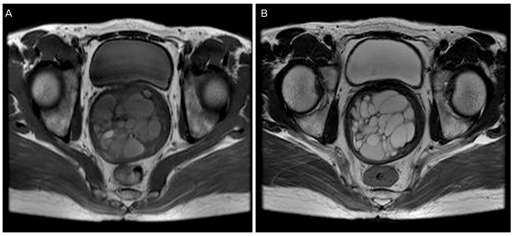
Fig. 1 (A) T1-weighted axial image of the pelvis showing a multiloculated cystic lesion not invading the cervical stroma with high-signal intensity. (B) T2-weighted axial image of the pelvis showing a multiloculated cystic lesion not invading the cervical stroma with high-signal intensity.
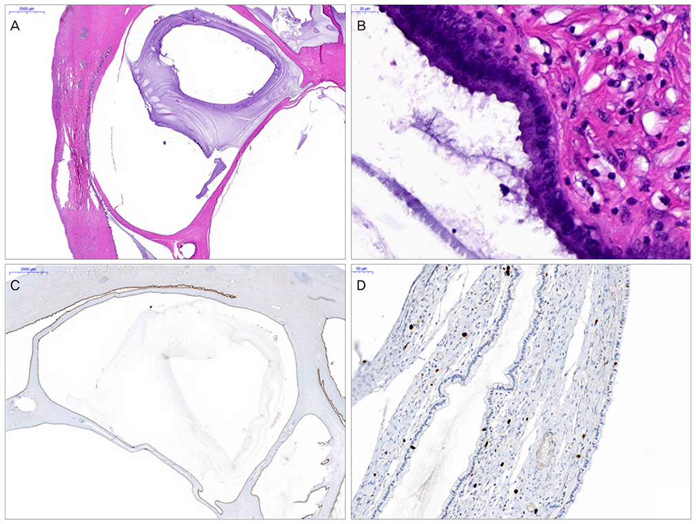
Fig. 2 (A) The cervix shows several deep-seated cystic spaces. The cysts are filled with mucin (H&E, ×20). (B) The cystic wall is lined by single layer of mucinous columnar epithelium (H&E, ×800). (C) The lining cells of the cyst show diffuse cytokeratin 7 expression (immunohistochemistry for cytokeratin 7, ×20). (D) The cystic wall shows a low Ki 67 labeling index (immunohistochemistry for Ki 67, ×400).
Reference
-
1. Batt RE, Yeh J. Mullerianosis: four developmental (embryonic) mullerian diseases. Reprod Sci. 2013; 20:1030–1037.2. Mobarki M, Karpathiou G, Forest F, Corsini T, Peoc’h M. Endocervicosis of the uterine cervix. Int J Gynecol Pathol. 2016; 35:475–477.3. Cheah PL, Looi LM, Lee GE, Teoh KH, Mun KS, Nazarina AR. Unusual finding of endocervical-like mucinous epithelium in continuity with urothelium in endocervicosis of the urinary bladder. Diagn Pathol. 2011; 6:56.4. McCluggage WG, Price JH, Dobbs SP. Primary adenocarcinoma of the vagina arising in endocervicosis. Int J Gynecol Pathol. 2001; 20:399–402.5. Olivia Vella JE, Nair N, Ferryman SR, Athavale R, Latthe P, Hirschowitz L. Mullerianosis of the urinary bladder. Int J Surg Pathol. 2011; 19:548–551.6. Mukonoweshuro P, McCluggage WG. Endocervicosis involving axillary lymph nodes: first case report. Int J Gynecol Pathol. 2014; 33:620–623.7. Slotta JE, Schaefer TJ, Walter B, Bohle RM, Schilling MK, Plusczyk T. Endocervicosis of the rectum. Int J Colorectal Dis. 2011; 26:683–684.8. Lim S, Kim JY, Park K, Kim BR, Ahn G. Mullerianosis of the mesosalpinx: a case report. Int J Gynecol Pathol. 2003; 22:209–212.9. Young RH, Clement PB. Endocervicosis involving the uterine cervix: a report of four cases of a benign process that may be confused with deeply invasive endocervical adenocarcinoma. Int J Gynecol Pathol. 2000; 19:322–328.10. Nucci MR. Pseudoneoplastic glandular lesions of the uterine cervix: a selective review. Int J Gynecol Pathol. 2014; 33:330–338.11. Eskridge MR, Rovner ES, Payne KD, Workman RB, Curry NS. MRI of endocervicosis: an unusual cause of clustered periurethral cystic masses involving the bladder. AJR Am J Roentgenol. 2007; 188:W147–W149.12. Castan Senar A, Pano B, Saco A, Nicolau C. Magnetic resonance imaging of adenoma malignum of the uterine cervix with pathologic correlation: a case report. Radiol Case Rep. 2016; 11:323–327.13. Park SB, Lee JH, Lee YH, Song MJ, Choi HJ. Multilocular cystic lesions in the uterine cervix: broad spectrum of imaging features and pathologic correlation. AJR Am J Roentgenol. 2010; 195:517–523.14. Kulkarni M, Hastak M, Chatterjee P. Adenoma malignum of the cervix: MR imaging appearance. Research. 2014; 1:635.15. Gong L, Zhang WD, Liu XY, Han XJ, Yao L, Zhu SJ, et al. Clonal status and clinicopathological observation of cervical minimal deviation adenocarcinoma. Diagn Pathol. 2010; 5:25.
- Full Text Links
-
- Actions
-
Cited
- CITED
-
- Close
- Share
-
- Similar articles
-
- Endocervicosis presenting as abdominal wall mass
- Muellerianosis of the urinary bladder, endocervicosis type: a case report
- Nursing Philosophy: Rethinking Nurses' Moral Dilemma and Self-cultivation from the Perspectives of Foucauldian Notions
- Biomechanics of Cervical Spine
- The Diagnostic Dilemma of Follicular Cholangitis Resembling Hilar Cholangiocarcinoma
