Successful Treatment of Internal Iliac Vein Rupture During Revisional Anterior Lumbar Spinal Surgery Using a Tack: Case Report
- Affiliations
-
- 1Department of Orthopaedic Surgery, Daegu Catholic University Medical Center, Daegu, Korea. bong@cu.ac.kr
- 2Department of General Surgery, Daegu Catholic University Medical Center, Daegu, Korea.
- KMID: 2385648
- DOI: http://doi.org/10.4184/jkss.2017.24.2.115
Abstract
- STUDY DESIGN: Case report.
OBJECTIVES
To report a rare case in which a tack was used to control bleeding due to a torn iliac vein during revisional anterior spine surgery. SUMMARY OF LITERATURE REVIEW: During anterior lumbar surgery, bleeding following a vascular injury is possible to control and reparable in most cases. During revisional anterior lumbar surgery, however, there are irreparable cases of bleeding as well. In some cases, it can threaten the patient's life.
MATERIALS AND METHODS
A 56-year-old man suffered from potentially fatal bleeding following iliac vein rupture during revisional anterior lumbar surgery. Primary vascular closure was impossible due to severe adhesion. We attempted to stop the venous bleeding with a tack, as an alternative treatment. The potentially fatal bleeding was controlled and the patient's vital signs stabilized after hemostasis by the tack.
RESULTS
Hemostasis using the tack saved the patient's life without any rebleeding.
CONCLUSIONS
During revisional anterior lumbar surgery, bleeding following an iliac vein rupture can be controlled by a tack in cases that are irreparable due to severe adhesion.
Keyword
MeSH Terms
Figure
-
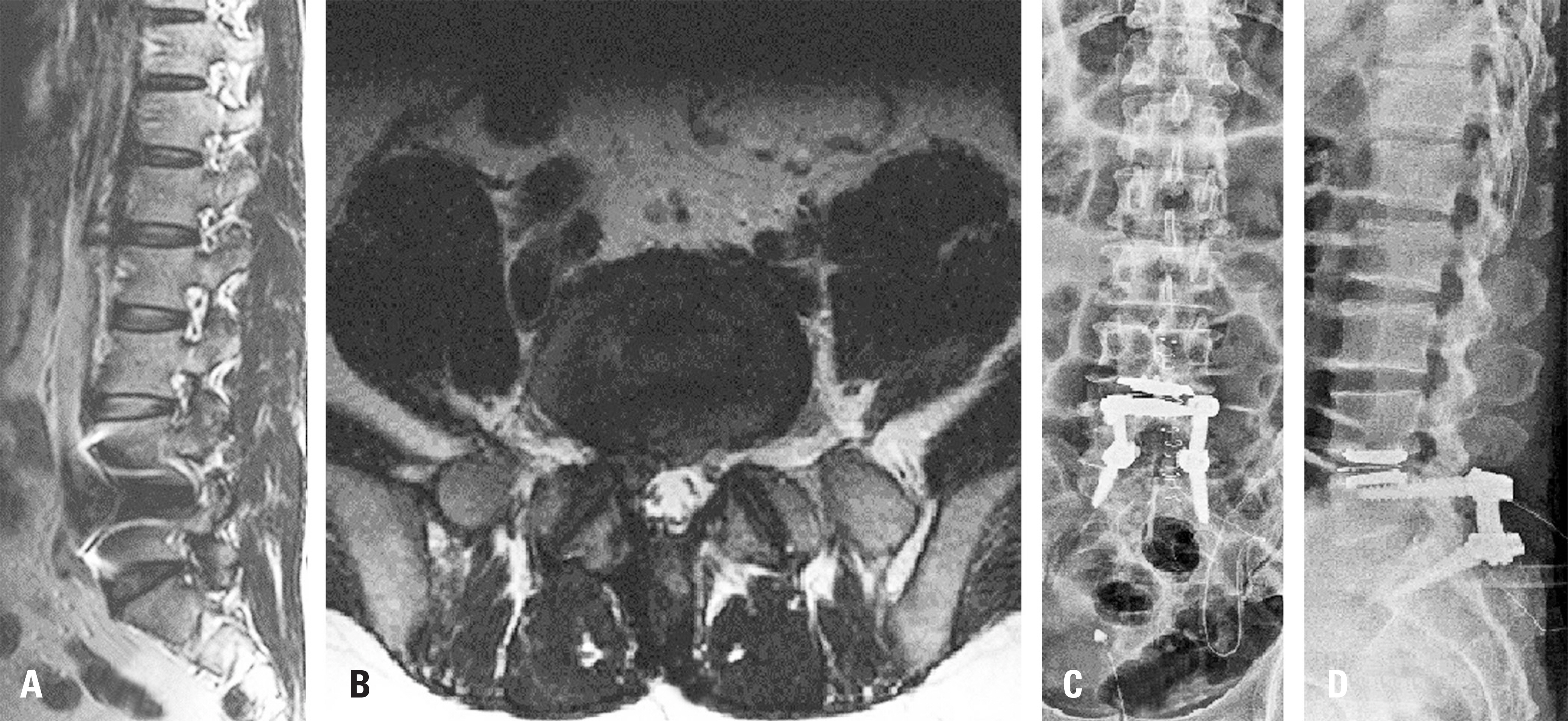
Fig. 1. (A) A sagittal image of the lumbar spine and (B) an axial image at L5-S1 show a left herniated disc in preoperative magnetic resonance imaging. (C) Coronal images and (D) sagittal images show decompressed lamina and posterior interbody fusion in postoperative imaging.
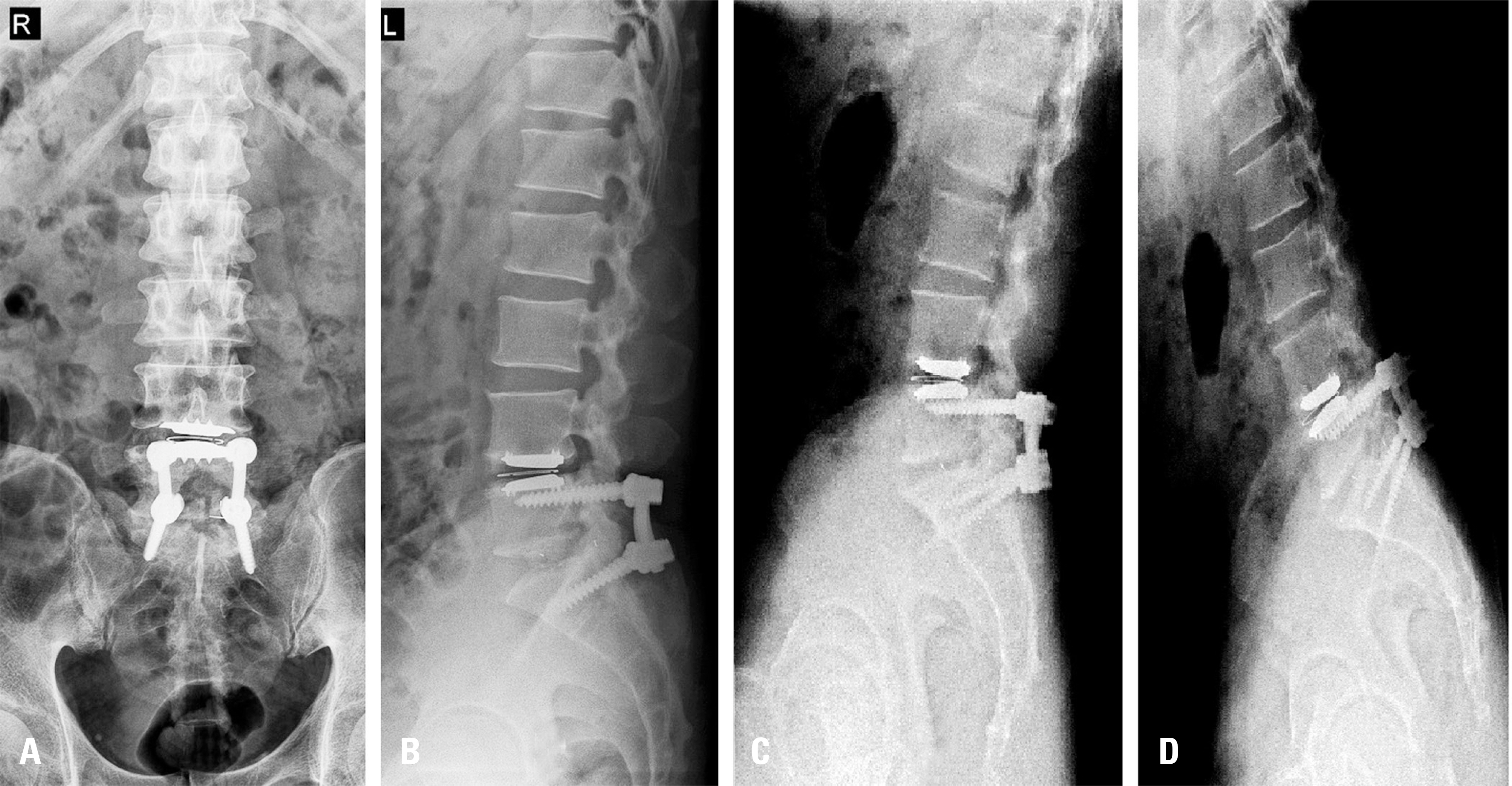
Fig. 2. (A) A coronal image, (B) a sagittal image, (C) an extension image, and (D) a flexion image show a displaced cage and non-fused findings at the L5-S1 level 1 year after the operation.

Fig. 3. A sagittal image of the lumbar spine shows a displaced cage and non-fused findings at the L5-S1 level in computed tomography.
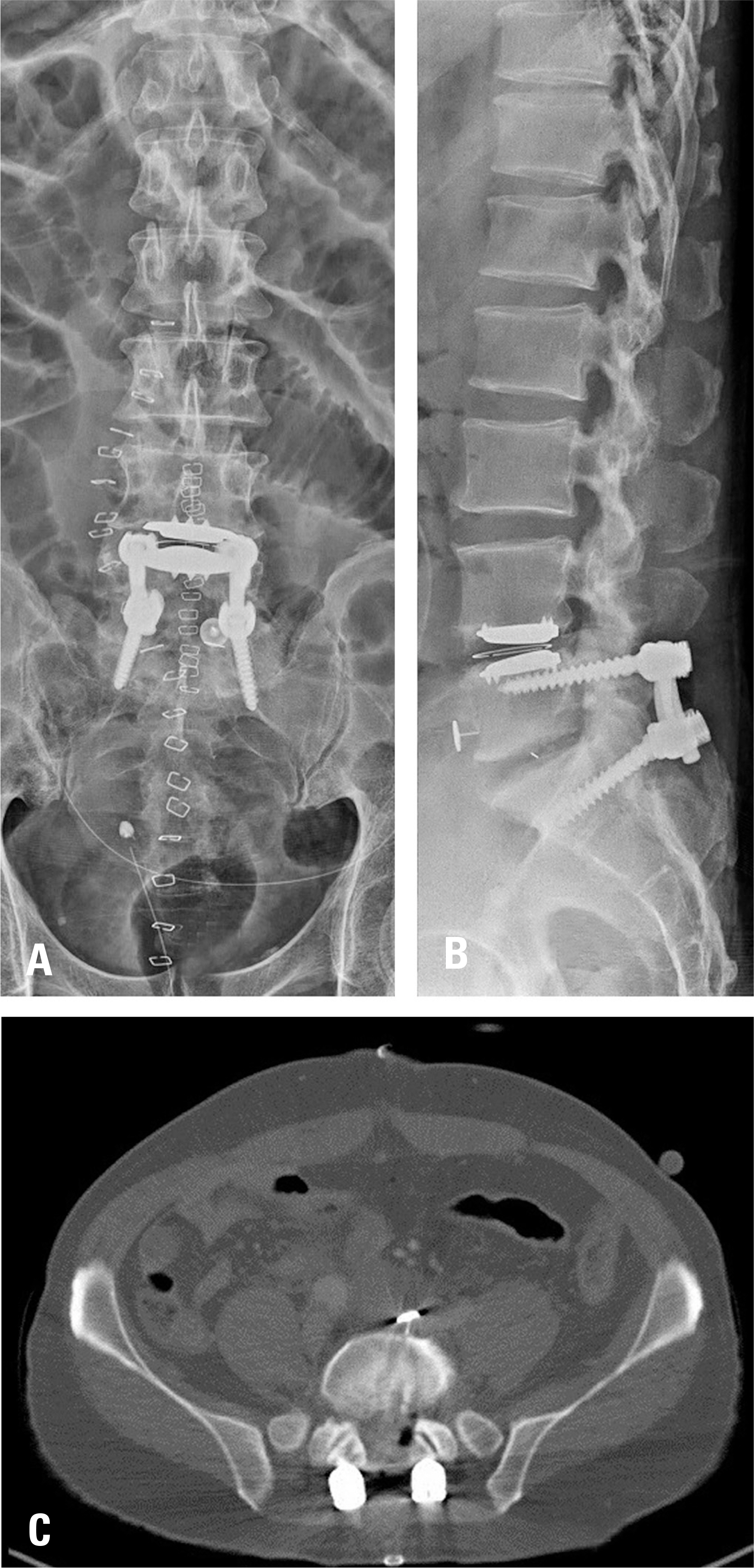
Fig. 4. The tack is shown maintaining its position well in (A) a coronal image and (B) a sagittal image obtained 3 days after the operation. Axial computed tomography images show acute venous thrombotic occlusion in images obtained 7 days postoperatively.
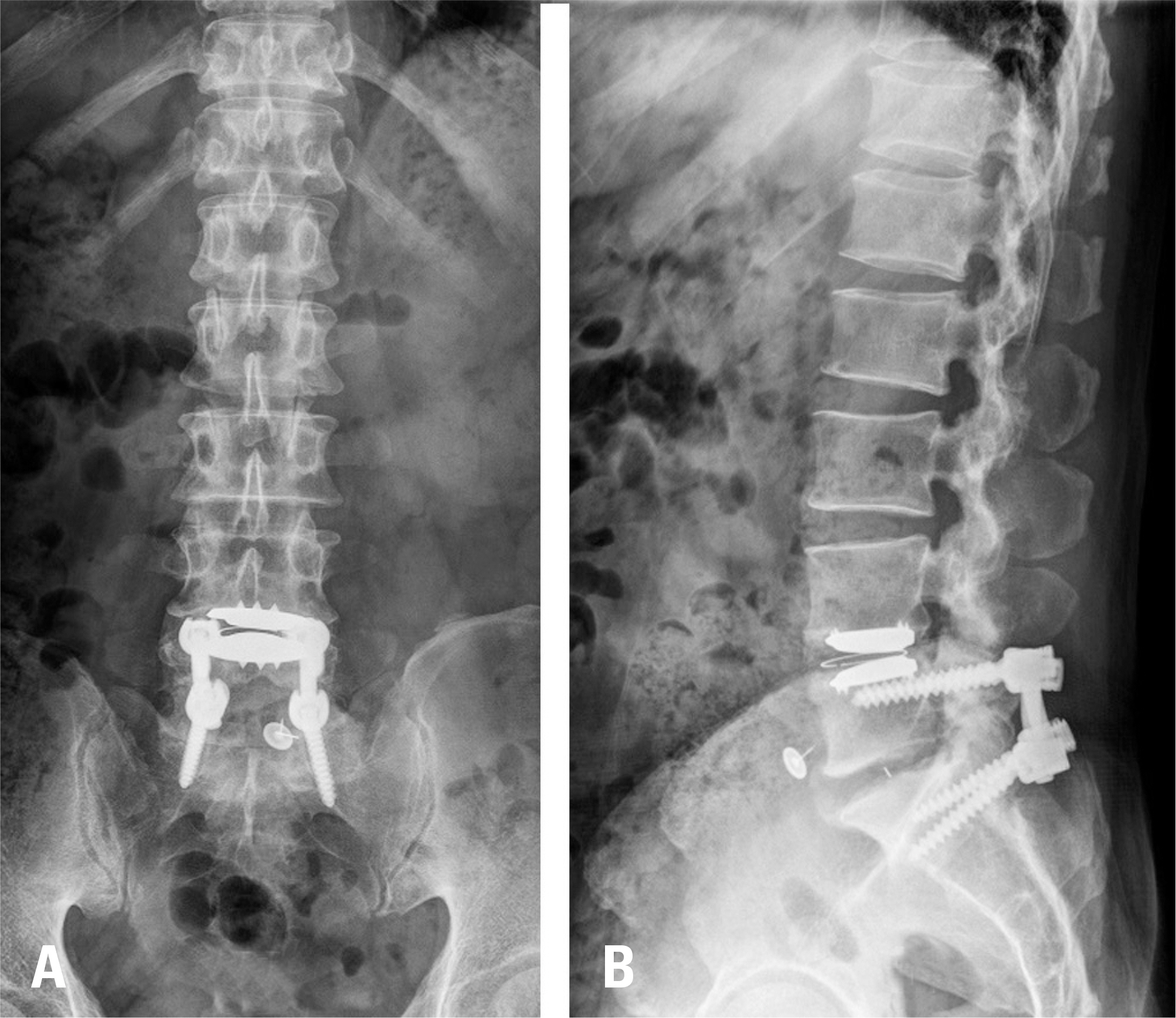
Fig. 5. (A) A coronal image and (B) a sagittal image obtained 1 year after the operation show the tack maintaining its position well.
Reference
-
1. Brau SA, Delamarter RB, Schiffman ML, et al. Vascular injury during anterior lumbar surgery. Spine (Phila Pa 1976). 2004; 4:409–12.2. Oskouian RJ, Johnson JP. Vascular complications in anterior thoracolumbar spinal reconstruction. J Neurosurg. 2002; 96:1–5.
Article3. Nguyen HV, Akbarnia BA, van Dam BE, et al. Anterior exposure of the spine for removal of lumbar interbody de-vices and implants. Spine (Phila Pa 1976). 2006; 31:2449–53.
Article4. Flouzat-Lachaniette CH, Delblond W, Poignard A, et al. Analysis of intraoperative difficulties and management of operative complications in revision anterior exposure of the lumbar spine: a report of 25 consecutive cases. Eur Spine J. 2013; 22:766–74.
Article5. Patel AA, Brodke DS, Pimenta L, et al. Revision strategies in lumbar total disc arthroplasty. Spine (Phila Pa 1976). 2008; 33:1276–83.
Article6. Schwender JD, Casnellie MT, Perra JH, et al. Perioperative complications in revision anterior lumbar spine surgery: incidence and risk factors. Spine (Phila Pa 1976). 2009; 34:87–90.7. Silvestre C, Mac-Thiong JM, Hilmi R, et al. Complications and morbidities of mini-open anterior retroperitoneal lumbar interbody fusion: Oblique lumbar interbody fusion in 179 patients. Asian Spine J. 2012; 6:89–97.
Article8. Chiriano J, Abou-Zamzam AM Jr, Urayeneza O, et al. The role of the vascular surgeon in anterior retroperitoneal spine exposure: preservation of open surgical training. J Vasc Surg. 2009; 50:148–51.
Article9. Ariyoshi D, Sano S, Kawamura N. Inferior vena cava injury caused by an anteriorly migrated cage resulting in ligation: case report. J Neurosurg Spine. 2016; 24:409–12.
Article10. Zahradnik V, Kashyap VS. Alternative management of iliac vein injury during anterior lumbar spine exposure. Ann Vasc Surg. 2012; 26:E15–8.
Article
- Full Text Links
-
- Actions
-
Cited
- CITED
-
- Close
- Share
-
- Similar articles
-
- Spontaneous left external iliac vein rupture
- Dangerous twisted communications between external and internal iliac veins which might rupture during catheterization
- Spontaneous Rupture of the Left External Iliac Vein
- Spontaneous Rupture of Iliac Vein: A case report
- Lumbar Epidural Venography in the Diagnosis of Lumbar Disc Herniation
