Dangerous twisted communications between external and internal iliac veins which might rupture during catheterization
- Affiliations
-
- 1Department of Anatomy, Melaka Manipal Medical College (Manipal Campus), Manipal Academy of Higher Education, Manipal, India. nayaksathish@gmail.com
- KMID: 2430201
- DOI: http://doi.org/10.5115/acb.2018.51.4.309
Abstract
- In this report, four unusual communications between external and internal iliac veins of the left side have been presented. The lowest communication was the narrowest measuring about 2 mm in diameter, the second measured 6 mm, the third had a diameter of 7 mm and the last communication measured 5 mm in breadth. The upper three communications were twisted in a helical manner. The internal iliac vein had its normal tributaries except that the iliolumbar vein drained into the external iliac vein at the level of the third communication. The external iliac vein was slightly dilated just below the level of lowest communication.
Keyword
Figure
-
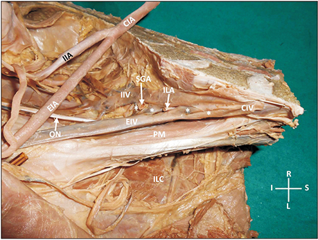
Fig. 1 Photograph of dissection of left iliac vessels in the left hemipelvis. Asterisks are showing four unusual communications between the external and internal iliac veins. CIA, common iliac artery; CIV, common iliac vein; EIA, external iliac artery; EIV, external iliac vein; IIA, internal iliac artery; IIV, internal iliac vein; ILA, iliolumbar artery; ILC, iliacus; ON, obturator nerve; PM, psoas major; SGA, superior gluteal artery.

Fig. 2 Photograph of dissection of left iliac vessels after their removal from the pelvis. CIV, common iliac vein; EIV, external iliac vein; IIV, internal iliac vein; ILV, iliolumbar vein; 1, 2, 3, and 4, the unusual communications between external and internal iliac veins. Note the twisted nature of these communications.
Reference
-
1. Yahyayev A, Bulakci M, Yilmaz E, Ucar A, Sayin OA, Yekeler E. Absence of the right iliac vein and an unusual connection between both common femoral veins. Phlebology. 2013; 28:162–164.
Article2. Onkar D, Onkar P, Mitra K. Isolated bilateral external iliac vein aplasia. Surg Radiol Anat. 2013; 35:85–87.
Article3. Hayashi S, Naito M, Yakura T, Kumazaki T, Itoh M, Nakano T. A case of an additional right external iliac vein surrounding the right external iliac artery and lacking the right common iliac vein. Anat Sci Int. 2016; 91:106–109.
Article4. Kuma S, Ishida M, Nakamura Y, Okazaki J. Prearterial external iliac vein as a rare anomaly of the iliac vein. Ann Vasc Surg. 2015; 29:836.e15–836.e17.
Article5. Djedovic G, Putz D. Case report: description of a venous annulus of the external iliac vein. Ann Anat. 2006; 188:451–453.
Article6. Surucu HS, Erbil KM, Tastan C, Yener N. Anomalous veins of the retroperitoneum: clinical considerations. Surg Radiol Anat. 2001; 23:443–445.
Article7. Shin M, Lee JB, Park SB, Park HJ, Kim YS. Multidetector computed tomography of iliac vein variation: prevalence and classification. Surg Radiol Anat. 2015; 37:303–309.
Article8. Yamaguchi T, Miyamoto T, Yamauchi Y, Obayashi T. A case report of successful permanent pacemaker implantation via the iliac vein. J Arrhythm. 2016; 32:151–153.
Article9. Pinski SL, Bredikis AJ. Defibrillator implantation via the iliac vein. Pacing Clin Electrophysiol. 2000; 23:1315–1317.
Article10. Okano K, Oshima M, Suzuki Y. Hepatic venous outflow reconstruction using an external iliac vein graft for hepatic malignancies (with video). J Hepatobiliary Pancreat Sci. 2012; 19:85–90.
Article11. Raju S, Buck WJ, Crim W, Jayaraj A. Optimal sizing of iliac vein stents. Phlebology. 2018; 33:451–457.
Article
- Full Text Links
-
- Actions
-
Cited
- CITED
-
- Close
- Share
-
- Similar articles
-
- Spontaneous left external iliac vein rupture
- Roentgenographic Confirmation of Central Venous Catheter Tips through the External and Internal Jugular Veins in Children
- External iliac artery injury with posterior pelvic ring injury in Korea: a report of two cases
- Roentgenographic Confirmation of Central Venous Catheter Tips Through the Basilic and External Jugular Veins
- Spontaneous Rupture of the Left External Iliac Vein
