Investig Magn Reson Imaging.
2017 Jun;21(2):114-118. 10.13104/imri.2017.21.2.114.
Skull Base Dermoid Cyst in the Right Infratemporal Fossa Diagnosed Using the Dixon Technique: a Case Report and Review of Literature
- Affiliations
-
- 1Department of Radiology, Haeundae Paik Hospital, Inje University College of Medicine, Busan, Korea.
- 2Department of Radiology, Gyeongsang National University School of Medicine and Gyeongsang National University Changwon Hospital, Changwon, Korea. sartre81@gmail.com
- 3Department of Neurology, Gyeongsang National University School of Medicine and Gyeongsang National University Changwon Hospital, Changwon, Korea.
- 4Department of Pathology, Gyeongsang National University School of Medicine and Gyeongsang National University Changwon Hospital, Changwon, Korea.
- KMID: 2385610
- DOI: http://doi.org/10.13104/imri.2017.21.2.114
Abstract
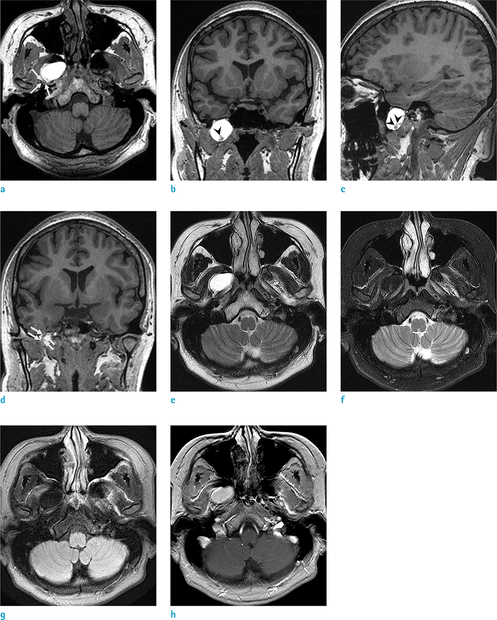
- Dermoid cysts are benign congenital tumors composed of keratinizing squamous epithelium and dermal derivatives. They account for less than 1% of all intracranial tumors and are rarely exhibited at the base of the skull. To the best of our knowledge, only one case report has presented computed tomography and conventional T1-weighted magnetic resonance (MR) findings that revealed an infratemporal dermoid cyst. In the present study, we report an unusual case of a dermoid cyst in the right infratemporal fossa, which was incidentally detected by MR imaging with the Dixon technique. This article also highlights the importance of meticulous radiological review and the usefulness of the Dixon technique in everyday clinical practice.
Figure
Reference
-
1. Guidetti B, Gagliardi FM. Epidermoid and dermoid cysts. Clinical evaluation and late surgical results. J Neurosurg. 1977; 47:12–18.2. Caldarelli M, Massimi L, Kondageski C, Di Rocco C. Intracranial midline dermoid and epidermoid cysts in children. J Neurosurg. 2004; 100:473–480.3. Watanabe K, Filomena CA, Nonaka Y, et al. Extradural dermoid cyst of the anterior infratemporal fossa. Case report. J Neurol Surg Rep. 2015; 76:e195–e199.4. Arseni C, Danaila L, Constantinescu AI, Carp N, Decu P. Cerebral dermoid tumours. Neurochirurgia (Stuttg). 1976; 19:104–114.5. North KN, Antony JH, Johnston IH. Dermoid of cavernous sinus resulting in isolated oculomotor nerve palsy. Pediatr Neurol. 1993; 9:221–223.6. Oursin C, Wetzel SG, Lyrer P, Bachli H, Stock KW. Ruptured intracranial dermoid cyst. J Neurosurg Sci. 1999; 43:217–220. discussion 220-211.7. Wilms G, Casselman J, Demaerel P, Plets C, De Haene I, Baert AL. CT and MRI of ruptured intracranial dermoids. Neuroradiology. 1991; 33:149–151.8. Rubin G, Scienza R, Pasqualin A, Rosta L, DaPian R. Craniocerebral epidermoids and dermoids. A review of 44 cases. Acta Neurochir (Wien). 1989; 97:1–16.9. Del Grande F, Santini F, Herzka DA, et al. Fat-suppression techniques for 3-T MR imaging of the musculoskeletal system. Radiographics. 2014; 34:217–233.10. Dixon WT. Simple proton spectroscopic imaging. Radiology. 1984; 153:189–194.
- Full Text Links
- Actions
-
Cited
- CITED
-
- Close
- Share
- Similar articles
-
- A Ruptured Dermoid Cyst of the Cavernous Sinus Extending into the Posterior Fossa
- Infratemporal Fossa Approach to Lesions in the Base of the Skull
- Surgical treatment of the lateral skull base tumor : type C infratemporal fossa approach
- Parapharyngeal Second Branchial Cleft Cyst Extending to the Skull Base: A Lateral Transcranial Infratemporal fossa Approach
- A Case of Endoscopic Drainage of Pterygoid Fossa Abscess Induced by Fungal Invasion


