Mucosal Incision and Forceps Biopsy for Reliable Tissue Sampling of Gastric Subepithelial Tumors
- Affiliations
-
- 1Department of Internal Medicine, Gangneung Asan Hospital, University of Ulsan College of Medicine, Gangneung, Korea. sangjin@gnah.co.kr
- KMID: 2383492
- DOI: http://doi.org/10.5946/ce.2015.094
Abstract
- BACKGROUND/AIMS
The diagnostic efficacy of current tissue sampling techniques for gastric subepithelial tumors (SETs) is limited. Better tissue sampling techniques are needed to improve pathological diagnosis. The aim of this study was to evaluate the safety and efficacy of a new technique, mucosal incision and forceps biopsy, for reliable tissue sampling of gastric SETs.
METHODS
This study enrolled 12 consecutive patients who underwent mucosal incision and forceps biopsy of gastric SETs between November 2011 and September 2014 at Gangneung Asan Hospital. The medical records of patients were reviewed retrospectively. The safety and diagnostic yield of this method were evaluated.
RESULTS
By performing mucosal incision and forceps biopsy, we were able to provide a definitive histological diagnosis for 11 out of 12 cases. The pathological diagnoses were leiomyoma (3/11), gastrointestinal stromal tumor (GIST; 2/11), lipoma (2/11), schwannoma (1/11), and ectopic pancreas (3/11). In cases of leiomyoma (n=3) and GIST (n=2), tissue samples were of sufficient size to allow immunohistochemical staining. In addition, the mitotic index was evaluated in two cases of GIST. There were no procedure-related complications.
CONCLUSIONS
Mucosal incision and forceps biopsy can be used as one of several methods to obtain adequate tissue samples from gastric SETs.
Keyword
MeSH Terms
Figure
-
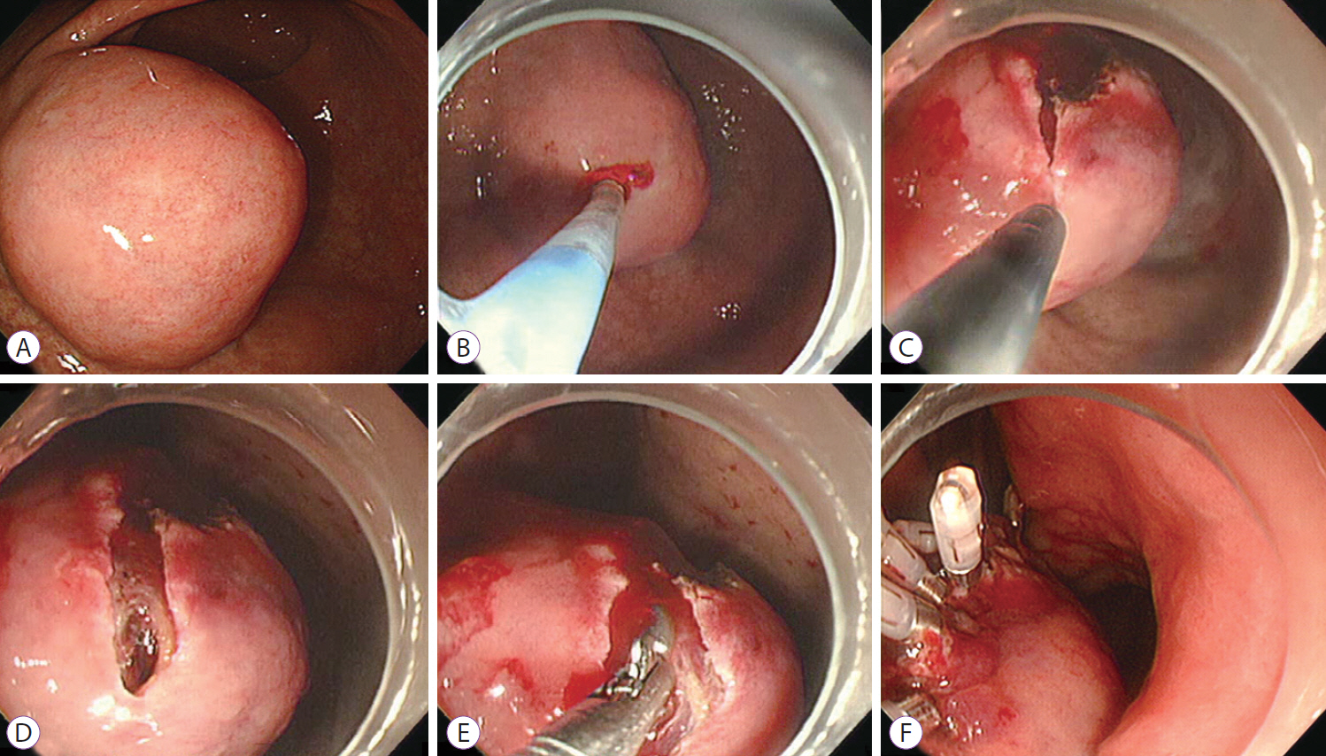
Fig. 1. Mucosal incision and forceps biopsy technique. (A) A 41-mm subepithelial tumor (SET) on the anterior wall of the angle of stomach. (B) Injection mixture of indigocarmine and glycerol in the SET. (C) Mucosal incision by using a hook knife. (D) Exposure of the SET through the incised mucosa. (E) Obtaining specmens by using conventional biopsy forceps. (F) Closure of the mucosal incisions with clips.

Fig. 2. Pathological findings for the biopsy specimen in a patient with schwannoma (Table 1, case 7). (A) Eight pieces of the biopsy specimen measuring 1 to 1.5 mm. (B) The biopsy specimen is composed of spindle cells (H&E stain, ×20). (C) The biopsy specimen showing reddish brown immunostaining, positive for S-100 (×20). (D) The biopsy specimen showing negative immunostaining for smooth muscle actin (SMA, ×20). (E) The biopsy specimen showing negative immunostaining for c-KIT (×20).
Cited by 3 articles
-
Comparison of the Diagnostic Ability of Endoscopic Ultrasonography and Abdominopelvic Computed Tomography in the Diagnosis of Gastric Subepithelial Tumors
Sang Yoon Kim, Ki-Nam Shim, Joo-Ho Lee, Ji Young Lim, Tae Oh Kim, A. Reum Choe, Chung Hyun Tae, Hye-Kyung Jung, Chang Mo Moon, Seong-Eun Kim, Sung-Ae Jung
Clin Endosc. 2019;52(6):565-573. doi: 10.5946/ce.2019.019.Diagnosis of Gastric Subepithelial Tumors Using Endoscopic Ultrasonography or Abdominopelvic Computed Tomography: Which is Better?
Eun Young Park, Gwang Ha Kim
Clin Endosc. 2019;52(6):519-520. doi: 10.5946/ce.2019.188.How Can We Obtain Tissue from a Subepithelial Lesion for Pathologic Diagnosis?
Eun Young Kim
Clin Endosc. 2017;50(1):6-7. doi: 10.5946/ce.2017.016.
Reference
-
1. Hedenbro JL, Ekelund M, Wetterberg P. Endoscopic diagnosis of submucosal gastric lesions. The results after routine endoscopy. Surg Endosc. 1991; 5:20–23.2. Polkowski M. Endoscopic ultrasound and endoscopic ultrasound-guided fine-needle biopsy for the diagnosis of malignant submucosal tumors. Endoscopy. 2005; 37:635–645.
Article3. Cantor MJ, Davila RE, Faigel DO. Yield of tissue sampling for subepithelial lesions evaluated by EUS: a comparison between forceps biopsies and endoscopic submucosal resection. Gastrointest Endosc. 2006; 64:29–34.
Article4. Ji JS, Lee BI, Choi KY, et al. Diagnostic yield of tissue sampling using a bite-on-bite technique for incidental subepithelial lesions. Korean J Intern Med. 2009; 24:101–105.
Article5. Jhala NC, Jhala D, Eltoum I, et al. Endoscopic ultrasound-guided fine-needle aspiration biopsy: a powerful tool to obtain samples from small lesions. Cancer. 2004; 102:239–246.
Article6. Hoda KM, Rodriguez SA, Faigel DO. EUS-guided sampling of suspected GI stromal tumors. Gastrointest Endosc. 2009; 69:1218–1223.
Article7. Sepe PS, Moparty B, Pitman MB, Saltzman JR, Brugge WR. EUS-guided FNA for the diagnosis of GI stromal cell tumors: sensitivity and cytologic yield. Gastrointest Endosc. 2009; 70:254–261.
Article8. Mekky MA, Yamao K, Sawaki A, et al. Diagnostic utility of EUS-guided FNA in patients with gastric submucosal tumors. Gastrointest Endosc. 2010; 71:913–919.
Article9. Fernandez-Esparrach G, Sendino O, Sole M, et al. Endoscopic ultrasound-guided fine-needle aspiration and trucut biopsy in the diagnosis of gastric stromal tumors: a randomized crossover study. Endoscopy. 2010; 42:292–299.
Article10. Lee CK, Chung IK, Lee SH, et al. Endoscopic partial resection with the unroofing technique for reliable tissue diagnosis of upper GI subepithelial tumors originating from the muscularis propria on EUS (with video). Gastrointest Endosc. 2010; 71:188–194.
Article11. Lee HL, Kwon OW, Lee KN, et al. Endoscopic histologic diagnosis of gastric GI submucosal tumors via the endoscopic submucosal dissection technique. Gastrointest Endosc. 2011; 74:693–695.
Article
- Full Text Links
-
- Actions
-
Cited
- CITED
-
- Close
- Share
-
- Similar articles
-
- Endoscopic Management of Gastric Subepithelial Tumor
- Advancements in the Diagnosis of Gastric Subepithelial Tumors
- Histological Comparison of Endoscopic Forceps Biopsy with Endoscopic Resection in Gastric Mucosal Elevated Lesion
- Using Forceps Biopsy after Small Submucosal Dissection in the Diagnosis of Gastric Subepithelial Tumors
- Tissue Acquisition in Gastric Epithelial Tumor Prior to Endoscopic Resection
