Hemoperitoneum from Spontaneous Rupture of a Metastatic Abdominal Lymph Node in Gallbladder Cancer: A Case Report
- Affiliations
-
- 1Department of Internal Medicine, Jeju National University School of Medicine, Jeju, Korea. suhmok@jejunu.ac.kr
- 2Department of Surgery, Jeju National University School of Medicine, Jeju, Korea.
- KMID: 2383436
- DOI: http://doi.org/10.4166/kjg.2017.69.1.79
Abstract
- Gallbladder (GB) cancer is asymptomatic in nature, making diagnosis and treatment difficult. The lymph node status is the strongest predictor of long-term survival for patients with GB cancer, and a complete removal of regional lymph nodes is important for patients undergoing radical resection of GB cancer. Unfortunately, lymph node metastases are common in the early stages of GB cancer. However, there have only been a few cases describing the symptoms or complications of metastatic lymph nodes in patients with GB cancer. Although hemoperitoneum caused by metastatic lymph nodes can occur with several cancers, it is very rare. To the best of our knowledge, hemoperitoneum from spontaneous ruptures of metastatic lymph nodes with GB cancer has not yet been reported. Herein, we describe such a case in a patient newly diagnosed with GB cancer.
Keyword
MeSH Terms
Figure
-
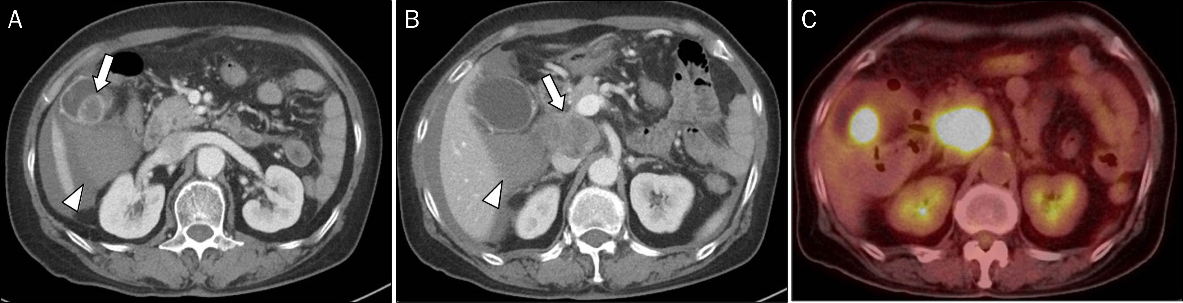
Fig. 1. Gallbladder cancer with metastatic portocaval lymph node. (A) A mass with a size of 2.5 cm in the gallbladder (arrow) and high density fluid collection are located near the gallbladder bed (arrowhead). (B) CT shows a soft-tissue mass with a size of approximately 5 cm at the portocaval space (arrow) and high density fluid around the mass (arrowhead). (C) A gallbladder polyp and a soft tissue mass show intense hypermetabolism on the PET scan finding.
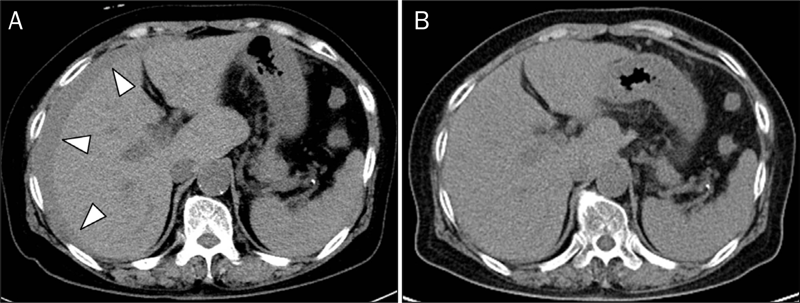
Fig. 2. Sequential changes of hemoperitoneum at the same level on coronal view of precontrast CT. (A) At admission, fluid collection is noted in the perihepatic space (arrowheads). (B) After 3 days, fluid collection is decreased without any interventions.
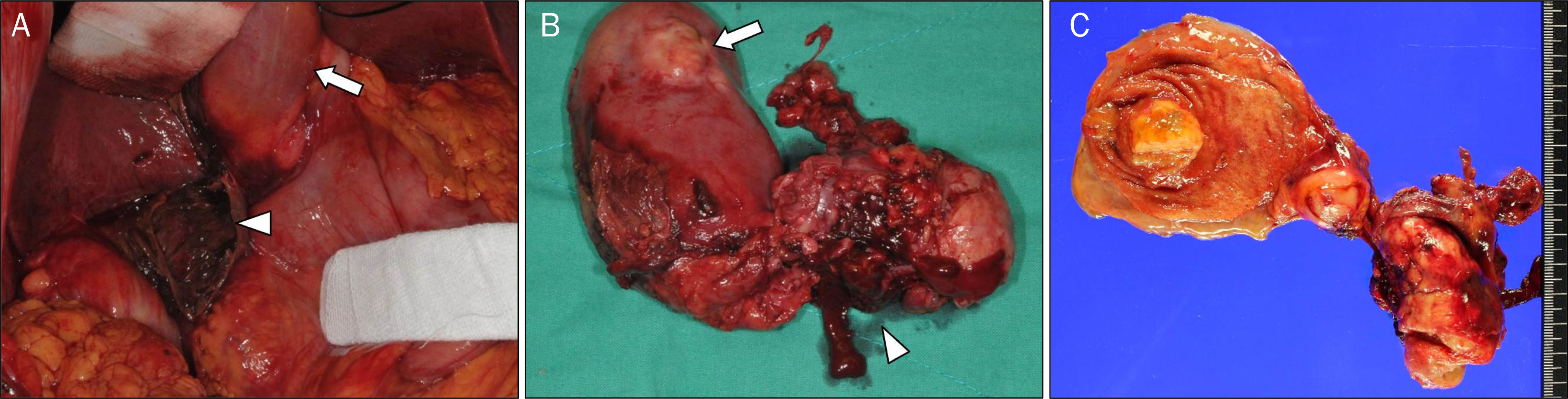
Fig. 3. Open cholecystectomy and lymph node dissection. (A) Hemoperitoneum is seen around the gallbladder (arrow) and ruptured lymph node (arrowhead). (B) Gallbladder wall is intact (arrow), but a metastatic lymph node is ruptured (arrowhead). (C) The surgically resected specimen shows 2.5 cm sized gallbladder cancer and 5cm sized ruptured metastatic lymph node.
Reference
-
References
1. Benoist S, Panis Y, Fagniez PL. Longterm results after curative resection for carcinoma of the gallbladder. French University Association for Surgical Research. Am J Surg. 1998; 175:118–122.2. Jensen EH, Abraham A, Jarosek S, et al. Lymph node evaluation is associated with improved survival after surgery for early stage gallbladder cancer. Surgery. 2009; 146:706–711. discussion 711–713.
Article3. Terzi C, Sökmen S, Seçkin S, Albayrak L, Uğ urlu M. Polypoid lesions of the gallbladder: report of 100 cases with special reference to operative indications. Surgery. 2000; 127:622–627.
Article4. Miller G, Jarnagin WR. Gallbladder carcinoma. Eur J Surg Oncol. 2008; 34:306–312.
Article5. Kondo S, Takada T, Miyazaki M, et al. Guidelines for the management of biliary tract and ampullary carcinomas: surgical treatment. J Hepatobiliary Pancreat Surg. 2008; 15:41–54.
Article6. Jensen EH, Abraham A, Habermann EB, et al. A critical analysis of the surgical management of early-stage gallbladder cancer in the United States. J Gastrointest Surg. 2009; 13:722–727.
Article7. Lucey BC, Varghese JC, Soto JA. Spontaneous hemoperitoneum: causes and significance. Curr Probl Diagn Radiol. 2005; 34:182–195.
Article8. Miyamoto M, Sudo T, Kuyama T. Spontaneous rupture of hepatocellular carcinoma: a review of 172 Japanese cases. Am J Gastroenterol. 1991; 86:67–71.9. Terada T, Takeuchi T, Hirano R, Nagata S, Kubota H, Honda S. Spontaneous rupture of peripancreatic lymph node with hepatocellular carcinoma metastasis: report of an autopsy case with massive peritoneal bleeding. Hepatol Res. 2003; 26:73–76.
Article10. Oh SY, Seo KW, Jegal Y, et al. Hemothorax caused by spontaneous rupture of a metastatic mediastinal lymph node in hepatocellular carcinoma: a case report. Korean J Intern Med. 2013; 28:622–625.
Article11. Seki S, Kitada T, Sakaguchi H, et al. Cardiac tamponade caused by spontaneous rupture of mediastinal lymph node metastasis of hepatocellular carcinoma. J Gastroenterol Hepatol. 2001; 16:702–704.
Article12. Moore K, Imbeault A, Roy G, Bolduc S. Massive hemorrhage from spontaneous rupture of a retroperitoneal lymph node in patient with metastatic mixed germ cell tumor. Urology. 2010; 76:159–161.
Article
- Full Text Links
-
- Actions
-
Cited
- CITED
-
- Close
- Share
-
- Similar articles
-
- Spontaneous Renal Rupture with Hemoperitoneum in a Patient on Continuous Ambulatory Peritoneal Dialysis
- A Case of Hemoperitoneum from A Spontaneous Venous Rupture Overlying Uterine Leiomyoma
- Spontaneous Rupture of Primary Angiosarcoma of the Spleen
- Spontaneous Rupture of the Intraperitoneal Metastatic Hepatocellular Carcinoma: a Case Report with Magnetic Resonance Imaging Findings
- Mucocutaneous Lymph Node Syndrome: Report of one case complicated by gallbladder hydrops and diagnosed by ultrasound
