Spontaneous Rupture of the Intraperitoneal Metastatic Hepatocellular Carcinoma: a Case Report with Magnetic Resonance Imaging Findings
- Affiliations
-
- 1Department of Radiology, Dankook University Hospital, Chungnam, Korea. deepva@hanmail.net
- KMID: 2421550
- DOI: http://doi.org/10.13104/imri.2018.22.3.177
Abstract
- Intraperitoneal metastatic hepatocellular carcinoma (HCC) is uncommon. Although rare, it can spontaneously rupture and cause hemoperitoneum similar to primary HCC in the liver. We present a case of intraperitoneal metastatic HCC that had spontaneously ruptured and appeared as an irregularly margined hemorrhagic mass with T1 high and T2 dark signal intensities on magnetic resonance imaging. Ruptured HCC is a life-threatening emergency with high mortality rate. Spontaneously ruptured intraperitoneal metastatic HCC should be considered if a patient with a history of HCC presents with acute abdomen, although rare.
MeSH Terms
Figure
-
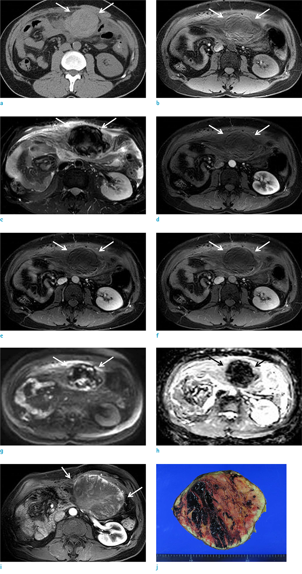
Fig. 1 A 40-year-old man with spontaneous rupture of intraperitoneal metastatic mass secondary to HCC. Axial non-enhanced CT scan (a) reveals an irregularly margined 8-cm high-density mass (arrows) in the omentum with hemoperitoneum. On MRI, the mass (arrows) reveals high signal intensity on pre-contrast T1-weighted image (b) and dark signal intensity on T2-weighted image (c). No significantly enhanced portion in the mass (arrows) was noted during dynamic enhancement study (d, arterial phase; e, portal phase; f, 3-min delayed phase). On DWI (b = 800 s/mm2) (g) and ADC map (h), the mass (arrows) does not reveal remarkable diffusion restriction. Follow-up MRI of the patient performed two months later revealed enlarged size, and more well-defined margin of the mass. Highly enhanced portion is well depicted at the periphery of the mass (arrows) on arterial phase image of dynamic enhancement study (i). Gross specimen (j) reveals a 15-cm metastatic HCC that appears to be a well-defined, firm, pink-tan, hemorrhagic mass.
Reference
-
1. van Malenstein H, van Pelt J, Verslype C. Molecular classification of hepatocellular carcinoma anno 2011. Eur J Cancer. 2011; 47:1789–1797.
Article2. Katyal S, Oliver JH 3rd, Peterson MS, Ferris JV, Carr BS, Baron RL. Extrahepatic metastases of hepatocellular carcinoma. Radiology. 2000; 216:698–703.
Article3. Okano J, Shiota G, Horie Y, et al. Rupture of metastatic nodule on the peritoneal surface secondary to hepatocellular carcinoma. Intern Med. 1996; 35:783–784.
Article4. Chen HW, Yang CF, Chao CC. Spontaneous rupture of peritoneal seeding hepatocellular carcinoma: report of two cases. J Acute Medicine. 2016; 6:64–66.
Article5. Tanaka A, Takeda R, Mukaihara S, et al. Treatment of ruptured hepatocellular carcinoma. Int J Clin Oncol. 2001; 6:291–295.
Article6. Spiliotis J, Nikolaou G, Kopanakis N, Vassiliadou D, Terra A, Efstathiou E. Hepatocellular carcinoma peritoneal metastasis: role of cytoreductive surgery and hyperthermic intraperitoneal chemotherapy (HIPEC). Gulf J Oncolog. 2017; 1:20–23.7. Casillas VJ, Amendola MA, Gascue A, Pinnar N, Levi JU, Perez JM. Imaging of nontraumatic hemorrhagic hepatic lesions. Radiographics. 2000; 20:367–378.
Article8. Zhu LX, Geng XP, Fan ST. Spontaneous rupture of hepatocellular carcinoma and vascular injury. Arch Surg. 2001; 136:682–687.
Article
- Full Text Links
-
- Actions
-
Cited
- CITED
-
- Close
- Share
-
- Similar articles
-
- Metastatic Omental Hepatocellular Carcinoma: Two Cases Report
- A Case of Hemoperitoneum Caused by Spontaneous Rupture of Metastatic Omental Hepatocellular Carcinoma
- Magnetic Resonance Imaging Finding of Metastatic Hepatocellular Carcinoma in Ovary: A Case Report
- Hemothorax Caused by Spontaneous Rupture of Hepatocellular Carcinoma in the Pleural Cavity: A Case Report
- A case of spontaneous hepatic rupture in a patient with primary hepatocellular carcinoma during the puerperium
