Primary Follicular Lymphoma in the Rectum Incidentally Found on Screening Colonoscopy
- Affiliations
-
- 1Department of Internal Medicine, Pusan National University Hospital, Pusan National University School of Medicine, Busan, Korea. gasong@pusan.ac.kr
- 2Department of Pathology, Pusan National University Hospital, Pusan National University School of Medicine, Busan, Korea.
- KMID: 2383424
- DOI: http://doi.org/10.4166/kjg.2017.69.2.139
Abstract
- The gastrointestinal tract is the most common site of extra-nodal non-Hodgkin lymphoma. However, the incidence of primary rectal lymphoma is extremely rare. Among the primary gastrointestinal lymphomas, follicular lymphoma has been described as a rare disease. It is difficult to diagnose rectal lymphoma due to its variable growth patterns and inadequate biopsies. Majority of patients with rectal lymphoma have non-specific symptoms or negative biopsies, often delaying the diagnosis. Our patient is a 62-year-old female. Two sessile and smooth subepithelial lesions with a yellowish normal mucosa were found on a screening colonoscopy. The initial mucosal biopsy finding was chronic inflammation, but we were highly suspicion of malignancy; we performed an endoscopic mucosal resection. Herein, we present a rare case of rectal follicular lymphoma diagnosed by endoscopic mucosal resection with a literature review.
Keyword
MeSH Terms
Figure
-
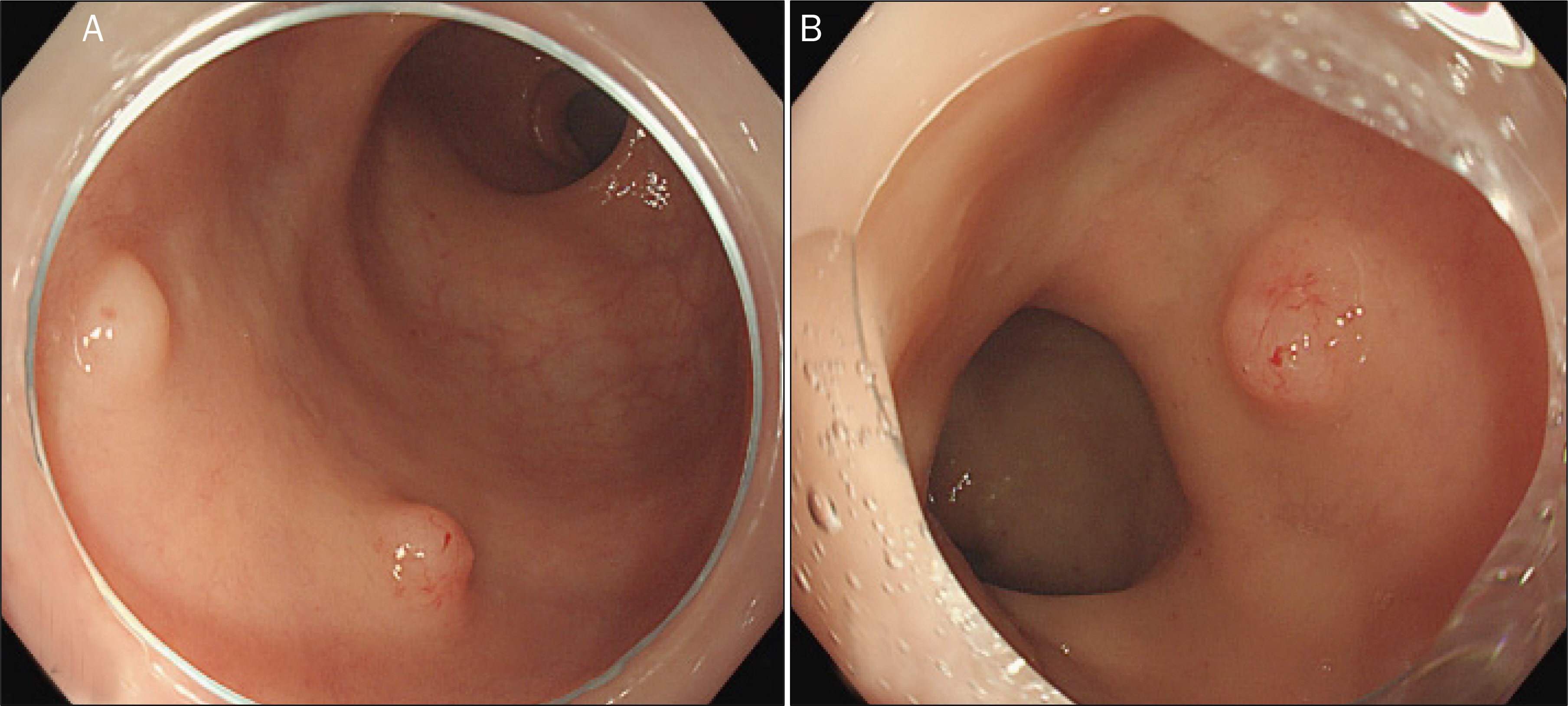
Fig. 1. Colonoscopic findings. (A) 4 mm subepithelial lesion on 3cm from anal verge, 3 mm subepitheliallesion on 2 cm from anal verge. (B) Smooth subepithelial lesion with yellowish colored normal mucosa on 3 cm from anal verge.
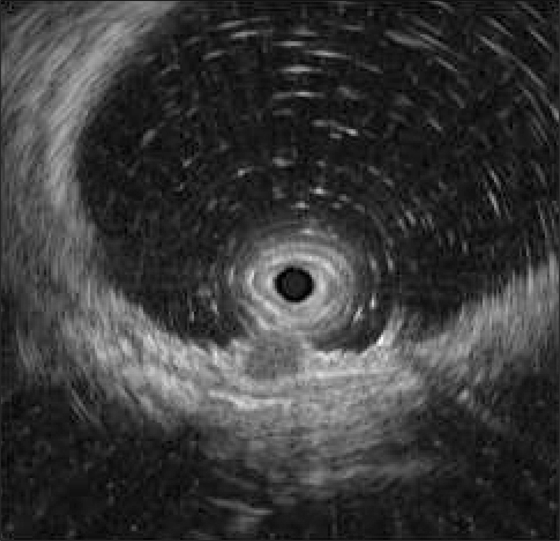
Fig. 2. EUS findings. 0.4×0.3 cm homogenous hypoechoic mass invaded second to third layer.

Fig. 3. Histologic findings. (A) Tumor is located in submucosa and invades into mucosa. The neoplastic follicles are poorly defined and show diffuse effacement (H&E, ×40). (B) Tumor cells are composed of centrocytes and centroblasts (H&E, ×400).

Fig. 4. Histologic findings. (A) Immunohistochemical staining for CD20. Tumor cells are positive for CD20 (×200). (B) Immunohistochemical staining for CD21 shows expanding follicular dendritic cell meshworks (×200).
Cited by 1 articles
-
Primary Colonic Follicular Lymphoma Presenting as Four Diminutive Sessile Polyps Found Incidentally During Colonoscopy
Sun Jin, Hyun Seok Lee, Ji Yun Jeong, Young Wook Jo
Clin Endosc. 2018;51(4):388-392. doi: 10.5946/ce.2017.114.
Reference
-
References
1. Rudders RA, Ross ME, DeLellis RA. Primary extranodal lymphoma: response to treatment and factors influencing prognosis. Cancer. 1978; 42:406–416.2. Koch P, del Valle F, Berdel WE, et al. Primary gastrointestinal non-Hodgkin's lymphoma: II. Combined surgical and conservative or conservative management only in localized gastric lymphoma–results of the prospective German Multicenter Study GIT NHL 01/92. J Clin Oncol. 2001; 19:3874–3883.
Article3. Fan CW, Changchien CR, Wang JY, et al. Primary colorectal lymphoma. Dis Colon Rectum. 2000; 43:1277–1282.
Article4. Yoshino T, Miyake K, Ichimura K, et al. Increased incidence of follicular lymphoma in the duodenum. Am J Surg Pathol. 2000; 24:688–693.
Article5. Dawson IM, Cornes JS, Morson BC. Primary malignant lymphoid tumours of the intestinal tract. Report of 37 cases with a study of factors influencing prognosis. Br J Surg. 1961; 49:80–89.
Article6. Koniaris LG, Drugas G, Katzman PJ, Salloum R. Management of gastrointestinal lymphoma. J Am Coll Surg. 2003; 197:127–141.
Article7. Shia J, Teruya-Feldstein J, Pan D, et al. Primary follicular lymphoma of the gastrointestinal tract: a clinical and pathologic study of 26 cases. Am J Surg Pathol. 2002; 26:216–224.8. Kwon BS, Kim CD, Park JY, et al. A case of primary follicular lymphoma arising in the rectum. Korean J Gastrointest Endosc. 2006; 33:285–288.9. Damaj G, Verkarre V, Delmer A, et al. Primary follicular lymphoma of the gastrointestinal tract: a study of 25 cases and a literature review. Ann Oncol. 2003; 14:623–629.
Article10. Iwamuro M, Okada H, Takata K, et al. Colorectal Manifestation of Follicular Lymphoma. Intern Med. 2016; 55:1–8.
Article11. West RB, Warnke RA, Natkunam Y. The usefulness of immunohistochemistry in the diagnosis of follicular lymphoma in bone marrow biopsy specimens. Am J Clin Pathol. 2002; 117:636–643.
Article12. Hoh CK, Glaspy J, Rosen P, et al. Whole-body FDG-PET imaging for staging of Hodgkin's disease and lymphoma. J Nucl Med. 1997; 38:343–348.13. Lapela M, Leskinen S, Minn HR, et al. Increased glucose metabolism in untreated non-Hodgkin's lymphoma: a study with positron emission tomography and fluorine-18-fluorodeoxyglucose. Blood. 1995; 86:3522–3527.14. Kahl BS, Yang DT. Follicular lymphoma: evolving therapeutic strategies. Blood. 2016; 127:2055–2063.
Article
- Full Text Links
-
- Actions
-
Cited
- CITED
-
- Close
- Share
-
- Similar articles
-
- Asymptomatic Small Bowel Lymphoma Discovered Incidentally Following Ileal Intubation During Screening Colonoscopy
- Primary Colonic Follicular Lymphoma Presenting as Four Diminutive Sessile Polyps Found Incidentally During Colonoscopy
- Mucosa-Associated Lymphoid-Tissue Lymphoma of the Cecum and Rectum: A Case Report
- A Case of Primary Follicular Lymphoma Arising in the Rectum
- A Case of Primary Rectal MALT Lymphoma Presented as Multiple Submucosal Tumors
