Ann Dermatol.
2011 Nov;23(4):490-492.
Milia-like Idiopathic Calcinosis Cutis Occurring in a Toddler Born as a Premature Baby
- Affiliations
-
- 1Department of Dermatology, School of Medicine and Medical Research Institute, Chungbuk National University, Cheongju, Korea. jyl@chungbuk.ac.kr
Abstract
- Milia-like idiopathic calcinosis cutis (MICC) is characterized by smooth, firm, whitish papules resembling milia. Histologically, it appears as a well-defined, round, basophilic nodule within the upper dermis. Although the etiology and treatment remain unclear, it may resolve spontaneously. Some cases have been associated with Down syndrome, and the mean age of MICC patients was 9.9 years old. Herein, we report a rare case of MICC that was not associated with Down syndrome. Noticeably, the patient, a toddler, was born as a premature baby and had an ischemic injury on the right foot at birth. However, the lesions appeared on both feet, including the non-injured left foot. Otherwise he was healthy. After a 21-month follow-up period, the lesions had almost disappeared without any treatment.
Keyword
Figure
-
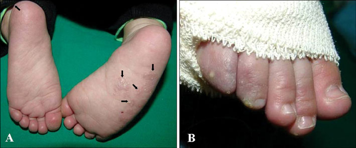
Fig. 1 (A) Multiple, firm, 1~4 mm sized, whitish papules on the right sole, left heel (arrows), and (B) right 5th toe. Morphologic changes on the right 4th and 5th toes may be caused by ischemic injury at birth.
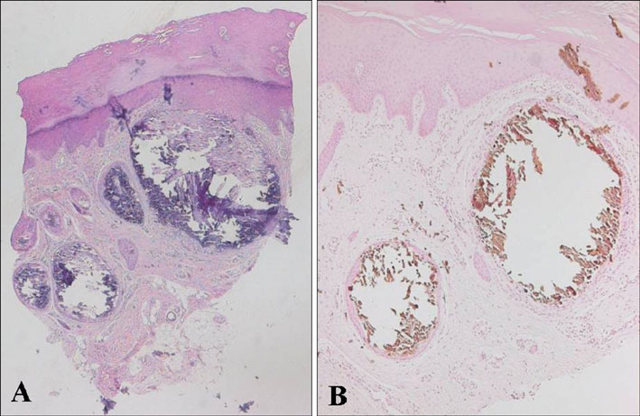
Fig. 2 (A) Several focal amorphous basophilic materials in the upper dermis and focal transepidermal elimination of the basophilic materials (H&E, ×40). (B) Von Kossa staining shows reaction of the basophilic materials, which were identified as calcium (Von Kossa stain, ×40).
Reference
-
1. Sano T, Tate S, Ishikawa C. A case of Down's syndrome associated with syringoma, milia, and subepidermal nodule. Jpn J Dermatol. 1978. 88:740.2. Menni S, Gualandri L, Boccardi D. Youngest case of idiopathic calcinosis cutis not associated with Down syndrome. Int J Dermatol. 2008. 47:870–871.
Article3. Choi WJ, Jung SJ, Seo YJ, Park EJ, Jo HJ, Kim KH, et al. A case of milia-like idiopathic calcinosis cutis that improved with tretinoin cream. Korean J Dermatol. 2008. 46:1640–1643.4. Kang H, Kim SE, Park K, Son SJ. A case of milia-like idiopathic calcinosis cutis in healthy boy. Korean J Dermatol. 2006. 44:868–870.5. Walsh JS, Fairley JA. Calcifying disorders of the skin. J Am Acad Dermatol. 1995. 33:693–706.
Article6. Walsh JS, Fairley JA. Wolff K, Goldsmith LA, Katz SI, Gilchrest BA, Paller AS, Leffell DJ, editors. Cutaneous mineralization and ossification. Fitzpatrick's dermatology in general medicine. 2008. 7th ed. New York: McGraw-Hill;1293–1297.7. Schepis C, Siragusa M, Palazzo R, Batolo D, Romano C. Milia-like idiopathic calcinosis cutis: an unusual dermatosis associated with Down syndrome. Br J Dermatol. 1996. 134:143–146.
Article8. Maroon M, Tyler W, Marks VJ. Calcinosis cutis associated with syringomas: a transepidermal elimination disorder in a patient with Down syndrome. J Am Acad Dermatol. 1990. 23:372–375.
Article9. Bécuwe C, Roth B, Villedieu MH, Chouvet B, Kanitakis J, Claudy A. Milia-like idiopathic calcinosis cutis. Pediatr Dermatol. 2004. 21:483–485.
Article10. Eng AM, Mandrea E. Peforating calcinosis cutis presenting as milia. J Cutan Pathol. 1981. 8:247–250.11. Ceder O, Roomans GM, Hösli P. Increased calcium content in cultured fibroblasts from trisomy patients: comparison with cystic fibrosis fibroblasts. Scan Electron Microsc. 1982. (Pt 2):723–730.12. Cambiaghi S, Imondi D, Gangi S, Vegni C. Fingertip calcinosis cutis. Cutis. 2000. 66:465–467.13. Schepis C, Siragusa M, Alberti A. Guess what! Milia-like idiopathic calcinosis cutis. Eur J Dermatol. 2000. 10:637–638.
- Full Text Links
-
- Actions
-
Cited
- CITED
-
- Close
- Share
-
- Similar articles
-
- A Case of Milia-like Idiopathic Calcinosis Cutis in an Elderly Person
- A Case of Milia-like Idiopathic Calcinosis Cutis That Improved with Tretinoin Cream
- A Case of Milia-like Idiopathic Calcinosis Cutis in Healthy Boy
- Two Cases of Milia-like Idiopathic Calcinosis Cutis Occurred in Infants
- A Case of Milia-like Idiopathic Calcinosis Cutis and Periorbital Syringomas in Down Syndrome
