Obstet Gynecol Sci.
2017 Jan;60(1):124-128. 10.5468/ogs.2017.60.1.124.
Extrauterine epithelioid trophoblastic tumor in hysterectomized woman
- Affiliations
-
- 1Department of Obstetrics and Gynecology, Samsung Medical Center, Sungkyunkwan University School of Medicine, Seoul, Korea. garden.lee@samsung.com
- 2Department of Obstetrics and Gynecology, Pusan National University Yangsan Hospital, Pusan National University School of Medicine, Yangsan, Korea.
- 3Department of Pathology, Samsung Medical Center, Sungkyunkwan University School of Medicine, Seoul, Korea.
- KMID: 2383220
- DOI: http://doi.org/10.5468/ogs.2017.60.1.124
Abstract
- Epithelioid trophoblastic tumor (ETT) is a very rare variant of gestational trophoblastic disease (GTD) which arises in reproductive age women with prior gestational history. Although abnormal vaginal bleeding is the most common symptom of ETT, there are no reported pathognomonic symptoms of ETT because of its rarity. ETT is similar to placental site trophoblastic tumor in terms of its slow growing characteristic and microscopic findings. Therefore, it could be misdiagnosed as placental site trophoblastic tumor or other types of GTD. Unlike other types of GTD, primary treatment of ETT is surgical resection because of its chemo-resistant nature. Accordingly, immunohistochemical staining is essential for accurate diagnosis and appropriate treatment. Here, we report a case of a 42-year-old hysterectomized woman with pelvic masses who suffered from abdominal pain. Through laparotomy, tumors were resected completely and they were diagnosed as ETT through immunohistochemical stain. This report provides more evidence about its clinical features, diagnosis, and treatment including a brief review of the literature.
Keyword
MeSH Terms
Figure
-
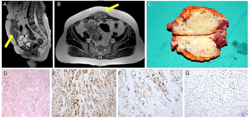
Fig. 1 Magnetic resonance imaging findings and pathologic evaluations of our patient. (A) Gross finding, midline lower abdominal wall mass with rectus abdominis muscle (photo during operation). (B,C) About 9-cm-sized midline lower abdominal wall soft tissue lesion with low signal intensity in T2W1 image on magnetic resonance imaging (arrowed). (B) Sagittal view and (C) transverse view. (D) The tumor was composed of nests of epithelioid cells with necrotic debris and peritumoral hyaline-like material (H&E, ×20). (E) Positive cytoplasmic staining for β-human chorionic gonadotropin (×100). (F) Positive cytoplasmic staining for inhibin-α (×100). (G) Positive nuclear staining for p63 (×100).
Reference
-
1. Shih IM, Kurman RJ. Epithelioid trophoblastic tumor: a neoplasm distinct from choriocarcinoma and placental site trophoblastic tumor simulating carcinoma. Am J Surg Pathol. 1998; 22:1393–1403.2. Lurain JR. Gestational trophoblastic disease I: epidemiology, pathology, clinical presentation and diagnosis of gestational trophoblastic disease, and management of hydatidiform mole. Am J Obstet Gynecol. 2010; 203:531–539.3. Lurain JR. Gestational trophoblastic disease II: classification and management of gestational trophoblastic neoplasia. Am J Obstet Gynecol. 2011; 204:11–18.4. Davis MR, Howitt BE, Quade BJ, Crum CP, Horowitz NS, Goldstein DP, et al. Epithelioid trophoblastic tumor: a single institution case series at the New England Trophoblastic Disease Center. Gynecol Oncol. 2015; 137:456–461.5. Scott EM, Smith AL, Desouki MM, Olawaiye AB. Epithelioid trophoblastic tumor: a case report and review of the literature. Case Rep Obstet Gynecol. 2012; 2012:862472.6. Palmer JE, Macdonald M, Wells M, Hancock BW, Tidy JA. Epithelioid trophoblastic tumor: a review of the literature. J Reprod Med. 2008; 53:465–475.7. Moutte A, Doret M, Hajri T, Peyron N, Chateau F, Massardier J, et al. Placental site and epithelioid trophoblastic tumours: diagnostic pitfalls. Gynecol Oncol. 2013; 128:568–572.8. Kim JY, An S, Jang SJ, Kim HR. Extrauterine epithelioid trophoblastic tumor of lung in a 35-year-old woman. Korean J Thorac Cardiovasc Surg. 2013; 46:471–474.9. Ahn HY, Hoseok I, Lee CH, Jung YJ, Shin NR, Kim KH, et al. Pulmonary mass diagnosed as extrauterine epithelioid trophoblastic tumor. Thorac Cardiovasc Surg. 2013; 61:97–100.10. Lewin SN, Aghajanian C, Moreira AL, Soslow RA. Extrauterine epithelioid trophoblastic tumors presenting as primary lung carcinomas: morphologic and immunohistochemical features to resolve a diagnostic dilemma. Am J Surg Pathol. 2009; 33:1809–1814.11. Li J, Shi Y, Wan X, Qian H, Zhou C, Chen X. Epithelioid trophoblastic tumor: a clinicopathological and immunohistochemical study of seven cases. Med Oncol. 2011; 28:294–299.12. Zhang X, Lu W, Lu B. Epithelioid trophoblastic tumor: an outcome-based literature review of 78 reported cases. Int J Gynecol Cancer. 2013; 23:1334–1338.
- Full Text Links
-
- Actions
-
Cited
- CITED
-
- Close
- Share
-
- Similar articles
-
- Extrauterine Epithelioid Trophoblastic Tumor of Lung in a 35-year-old Woman
- Epithelioid Trophoblastic Tumor in a Postmenopausal Woman: A Case Report
- Epitheilioid Trophoblastic Tumor of the Lung: A Case Report
- Epithelioid Trophoblastic Tumor: Clinicopathologic and Immunohistochemical Analysis of Three Cases
- Epithelioid trophoblastic tumor: A Case Report and Review of the Literature
