The Prognosis and Recovery of Aphasia Related to Stroke Lesion
- Affiliations
-
- 1Department of Rehabilitation Medicine, St. Vincent Hospital, College of Medicine, The Catholic University of Korea, Suwon, Korea. seonghoon@catholic.ac.kr
- KMID: 2382910
- DOI: http://doi.org/10.5535/arm.2016.40.5.786
Abstract
OBJECTIVE
To investigate the effects of specific brain lesions on prognosis and recovery of post-stroke aphasia, and to assess the characteristic pattern of recovery.
METHODS
Total of 15 subjects with first-ever, left hemisphere stroke, who were right handed, and who completed language assessment using the Korean version of the Western Aphasia Battery (K-WAB) at least twice during the subacute and chronic stages of stroke, were included. The brain lesions of the participants were evaluated using MRI-cron, SPM8, and Talairach Daemon software.
RESULTS
Subtraction of the lesion overlap map of the participants who showed more than 30% improvement in the aphasia quotient (AQ) by the time of their chronic stage (n=9) from the lesion overlap map of those who did not show more than 30% improvement in the AQ (n=6) revealed a strong relationship with Broca's area, inferior prefrontal gyrus, premotor cortex, and a less strong relationship with Wernicke's area and superior and middle temporal gyri. The culprit lesion related to poor prognosis, after grouping the subjects according to their AQ score in the chronic stage (a cut score of 50), revealed a strong relationship with Broca's area, superior temporal gyrus, and a less strong relationship with Wernicke's area, prefrontal cortex, and inferior frontal gyrus.
CONCLUSION
Brain lesions in the Broca's area, inferior prefrontal gyrus, and premotor cortex may be related to slow recovery of aphasia in patients with left hemisphere stroke. Furthermore, involvement of Broca's area and superior temporal gyrus may be associated with poor prognosis of post-stroke aphasia.
MeSH Terms
Figure
-
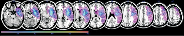
Fig. 1 Lesion overlap map for all participants (n=15). Color spectrum based on overlapping proportion (%).
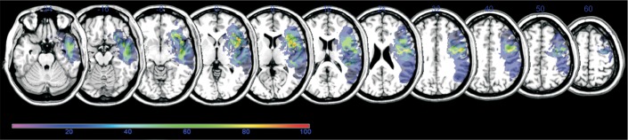
Fig. 2 Subtraction of overlay of participants with more than 30% AQ improvement (n=9) from that of those with less than 30% AQ improvement (n=6). Color spectrum based on overlapping proportion (%). AQ, aphasia quotient.
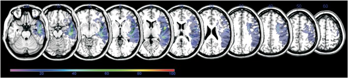
Fig. 3 Subtraction of overlay of participants with follow-up AQ more than 50 (n=6) from that of those with follow-up AQ less than 50 (n=9). Color spectrum based on overlapping proportion (%). AQ, aphasia quotient.
Cited by 1 articles
-
Changes in Language Function and Recovery-Related Prognostic Factors in First-Ever Left Hemispheric Ischemic Stroke
Kyung Ah Kim, Jung Soo Lee, Won Hyuk Chang, Deog Young Kim, Yong-Il Shin, Soo-Yeon Kim, Young Taek Kim, Sung Hyun Kang, Ji Yoo Choi, Yun-Hee Kim
Ann Rehabil Med. 2019;43(6):625-634. doi: 10.5535/arm.2019.43.6.625.
Reference
-
1. Hoffmann M, Chen R. The spectrum of aphasia subtypes and etiology in subacute stroke. J Stroke Cerebrovasc Dis. 2013; 22:1385–1392. PMID: 23680689.
Article2. Berthier ML, Pulvermüller F, Dávila G, Casares NG, Gutiérrez A. Drug therapy of post-stroke aphasia: a review of current evidence. Neuropsychol Rev. 2011; 21:302–317. PMID: 21845354.
Article3. Charidimou A, Kasselimis D, Varkanitsa M, Selai C, Potagas C, Evdokimidis I. Why is it difficult to predict language impairment and outcome in patients with aphasia after stroke. J Clin Neurol. 2014; 10:75–83. PMID: 24829592.
Article4. Cappa SF. The neural basis of aphasia rehabilitation: evidence from neuroimaging and neurostimulation. Neuropsychol Rehabil. 2011; 21:742–754. PMID: 22011017.
Article5. Crinion J, Holland AL, Copland DA, Thompson CK, Hillis AE. Neuroimaging in aphasia treatment research: quantifying brain lesions after stroke. Neuroimage. 2013; 73:208–214. PMID: 22846659.
Article6. Butler RA, Lambon Ralph MA, Woollams AM. Capturing multidimensionality in stroke aphasia: mapping principal behavioural components to neural structures. Brain. 2014; 137(Pt 12):3248–3266. PMID: 25348632.
Article7. Saur D, Hartwigsen G. Neurobiology of language recovery after stroke: lessons from neuroimaging studies. Arch Phys Med Rehabil. 2012; 93(1 Suppl):S15–S25. PMID: 22202187.
Article8. Lee KB, Kim JS, Hong BY, Kim YD, Hwang BY, Lim SH. The motor recovery related with brain lesion in patients with intracranial hemorrhage. Behav Neurol. 2015; 2015:258161. PMID: 25918457.
Article9. Kertesz A, Poole E. The aphasia quotient: the taxonomic approach to measurement of aphasic disability. Can J Neurol Sci. 1974; 1:7–16. PMID: 4434266.
Article10. Brett M, Leff AP, Rorden C, Ashburner J. Spatial normalization of brain images with focal lesions using cost function masking. Neuroimage. 2001; 14:486–500. PMID: 11467921.
Article11. Lancaster JL, Woldorff MG, Parsons LM, Liotti M, Freitas CS, Rainey L, et al. Automated Talairach atlas labels for functional brain mapping. Hum Brain Mapp. 2000; 10:120–131. PMID: 10912591.
Article12. Friederici AD, Ruschemeyer SA, Hahne A, Fiebach CJ. The role of left inferior frontal and superior temporal cortex in sentence comprehension: localizing syntactic and semantic processes. Cereb Cortex. 2003; 13:170–177. PMID: 12507948.
Article13. Gabrieli JD, Poldrack RA, Desmond JE. The role of left prefrontal cortex in language and memory. Proc Natl Acad Sci U S A. 1998; 95:906–913. PMID: 9448258.
Article14. Poldrack RA, Wagner AD, Prull MW, Desmond JE, Glover GH, Gabrieli JD. Functional specialization for semantic and phonological processing in the left inferior prefrontal cortex. Neuroimage. 1999; 10:15–35. PMID: 10385578.
Article15. Itabashi R, Nishio Y, Kataoka Y, Yazawa Y, Furui E, Matsuda M, et al. Damage to the left precentral gyrus is associated with apraxia of speech in acute stroke. Stroke. 2016; 47:31–36. PMID: 26645260.
Article16. Blank I, Balewski Z, Mahowald K, Fedorenko E. Syntactic processing is distributed across the language system. Neuroimage. 2016; 127:307–323. PMID: 26666896.
Article17. Meister IG, Wilson SM, Deblieck C, Wu AD, Iacoboni M. The essential role of premotor cortex in speech perception. Curr Biol. 2007; 17:1692–1696. PMID: 17900904.
Article18. Cohen YE, Theunissen F, Russ BE, Gill P. Acoustic features of rhesus vocalizations and their representation in the ventrolateral prefrontal cortex. J Neurophysiol. 2007; 97:1470–1484. PMID: 17135477.
Article19. Eickhoff SB, Heim S, Zilles K, Amunts K. A systems perspective on the effective connectivity of overt speech production. Philos Trans A Math Phys Eng Sci. 2009; 367:2399–2421. PMID: 19414462.
Article20. Ballard KJ, Tourville JA, Robin DA. Behavioral, computational, and neuroimaging studies of acquired apraxia of speech. Front Hum Neurosci. 2014; 8:892. PMID: 25404911.
Article21. Lee B, Pyun SB. Characteristics of cognitive impairment in patients with post-stroke aphasia. Ann Rehabil Med. 2014; 38:759–765. PMID: 25566474.
Article22. Seniow J, Litwin M, Lesniak M. The relationship between non-linguistic cognitive deficits and language recovery in patients with aphasia. J Neurol Sci. 2009; 283:91–94. PMID: 19268973.23. Yu ZZ, Jiang SJ, Bi S, Li J, Lei D, Sun LL. Relationship between linguistic functions and cognitive functions in a clinical study of Chinese patients with post-stroke aphasia. Chin Med J (Engl). 2013; 126:1252–1256. PMID: 23557554.24. Kertesz A, Harlock W, Coates R. Computer tomographic localization, lesion size, and prognosis in aphasia and nonverbal impairment. Brain Lang. 1979; 8:34–50. PMID: 476474.
Article25. Mazzocchi F, Vignolo LA. Localisation of lesions in aphasia: clinical-CT scan correlations in stroke patients. Cortex. 1979; 15:627–653. PMID: 95004.
Article26. Watila MM, Balarabe SA. Factors predicting poststroke aphasia recovery. J Neurol Sci. 2015; 352:12–18. PMID: 25888529.27. Heiss WD, Thiel A, Kessler J, Herholz K. Disturbance and recovery of language function: correlates in PET activation studies. Neuroimage. 2003; 20(Suppl 1):S42–S49. PMID: 14597295.
Article
- Full Text Links
-
- Actions
-
Cited
- CITED
-
- Close
- Share
-
- Similar articles
-
- Subcortical Aphasia in Stroke Patients
- Types, Severity and Prognostic Factors in Subcortical Aphasia
- Analysis of Aphasia Patients Resulting from Acute Ischemic Stroke Using Quantitative Methods of Aphasia Test
- Characteristics of Cognitive Impairment in Patients With Post-stroke Aphasia
- Predictors of Therapy Response in Chronic Aphasia: Building a Foundation for Personalized Aphasia Therapy
