Pityriasis Lichenoides-like Mycosis Fungoides: Clinical and Histologic Features and Response to Phototherapy
- Affiliations
-
- 1Department of Dermatology, Kosin University College of Medicine, Busan, Korea. ksderm77@unitel.co.kr
- 2Department of Internal Medicine, Kosin University College of Medicine, Busan, Korea.
- KMID: 2382876
- DOI: http://doi.org/10.5021/ad.2016.28.5.540
Abstract
- BACKGROUND
Pityriasis lichenoides (PL)-like skin lesions rarely appear as a specific manifestation of mycosis fungoides (MF).
OBJECTIVE
We investigated the clinicopathological features, immunophenotypes, and treatments of PL-like MF.
METHODS
This study included 15 patients with PL-like lesions selected from a population of 316 patients diagnosed with MF at one institution.
RESULTS
The patients were between 4 and 59 years of age. Four patients were older than 20 years of age. All of the patients had early-stage MF. In all patients, the atypical lymphocytic infiltrate had a perivascular distribution with epidermotropism. The CD4/CD8 ratio was <1 in 12 patients. Thirteen patients were treated with either narrowband ultraviolet B (NBUVB) or psoralen+ultraviolet A (PUVA), and all of them had complete responses.
CONCLUSION
PL-like MF appears to have a favorable prognosis and occurrence of this variant in adults is uncommon. MF should be suspected in the case of a PL-like skin eruption. Therefore, biopsy is required to confirm the diagnosis of PL-like MF, and NBUVB is a clinically effective treatment.
MeSH Terms
Figure
-
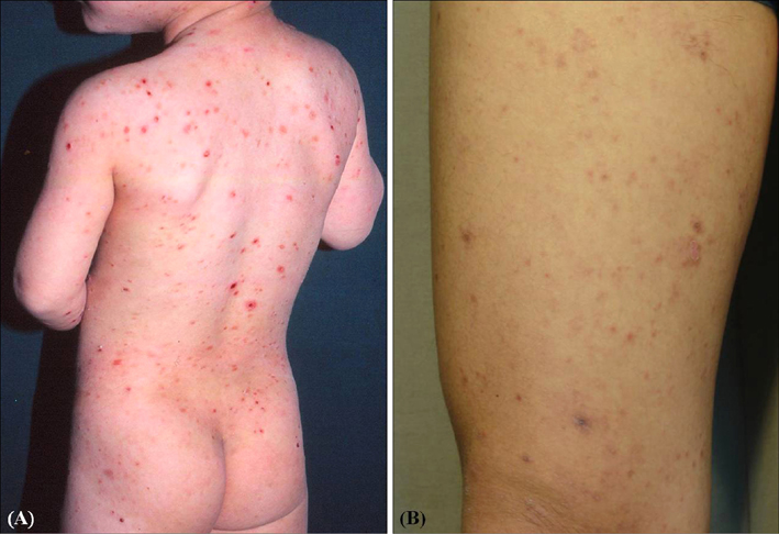
Fig. 1 (A) Generalized erythematous scaly discrete papules on the whole body (patient 1). (B) Brownish to erythematous crustered scaly papules (patient 6).
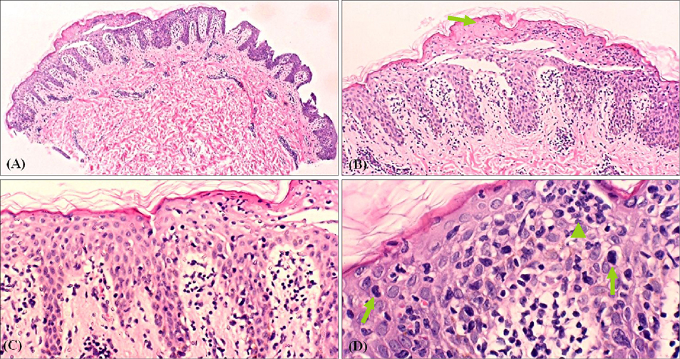
Fig. 2 (A) Biopsy specimens showing psoriasiform lichenoid infiltration (H&E, ×40). (B) Upper epidermal necrosis (arrow) and dermal papillae stuffed with lymphocytes (H&E, ×100). (C) Epidermotropism, "haloed" lymphocytes, and basal vacuolar change (H&E, ×200). (D) Intraepidermal lymphocytes that are larger than those in the dermis, hyperchromatic atypical cells (arrows), and Pautrier's microabscess (arrowhead) (H&E, ×400).
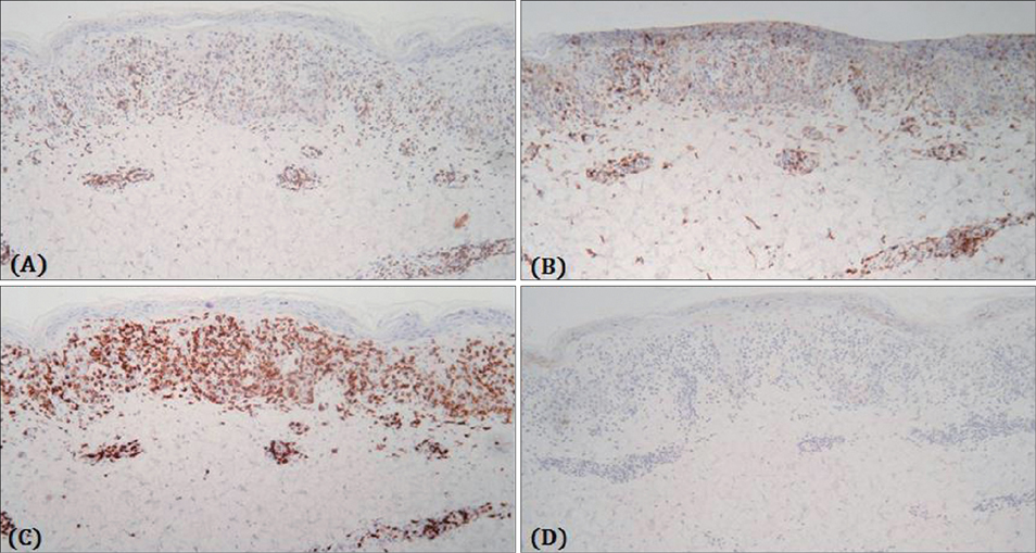
Fig. 3 Some atypical lymphocytes with positive immunohistochemical staining of (A) CD3, (B) CD4, (C) CD8, and negative staining of (D) CD30. CD8-positive T cells are predominant than CD4-positive T cells in the dermis, and CD4-positive T cells are predominant than CD8-positive T cells in the epidermis (×100) (patient 7).
Cited by 1 articles
-
Folliculotropic Mycosis Fungoides in 20 Korean Cases: Clinical and Histopathologic Features and Response to Ultraviolet A-1 and/or Photodynamic Therapy
Min Soo Jang, Ji Yun Jang, Jong Bin Park, Dong Young Kang, Jin Woo Lee, Taek Geun Lee, Hyun Hwangbo, Kee Suck Suh
Ann Dermatol. 2018;30(2):192-201. doi: 10.5021/ad.2018.30.2.192.
Reference
-
1. Jawed SI, Myskowski PL, Horwitz S, Moskowitz A, Querfeld C. Primary cutaneous T-cell lymphoma (mycosis fungoides and Sézary syndrome): part I. Diagnosis: clinical and histopathologic features and new molecular and biologic markers. J Am Acad Dermatol. 2014; 70:205.e1–205.e16.2. Ahn CS, ALSayyah A, Sangüeza OP. Mycosis fungoides: an updated review of clinicopathologic variants. Am J Dermatopathol. 2014; 36:933–948.3. Ko JW, Seong JY, Suh KS, Kim ST. Pityriasis lichenoides-like mycosis fungoides in children. Br J Dermatol. 2000; 142:347–352.
Article4. Olsen E, Vonderheid E, Pimpinelli N, Willemze R, Kim Y, Knobler R, et al. Revisions to the staging and classification of mycosis fungoides and Sezary syndrome: a proposal of the International Society for Cutaneous Lymphomas (ISCL) and the cutaneous lymphoma task force of the European Organization of Research and Treatment of Cancer (EORTC). Blood. 2007; 110:1713–1722.
Article5. Pimpinelli N, Olsen EA, Santucci M, Vonderheid E, Haeffner AC, Stevens S, et al. Defining early mycosis fungoides. J Am Acad Dermatol. 2005; 53:1053–1063.
Article6. de Unamuno Bustos B, Ferriols AP, Sánchez RB, Rabasco AG, Vela CG, Piris MA, et al. Adult pityriasis lichenoides-like mycosis fungoides: a clinical variant of mycosis fungoides. Int J Dermatol. 2014; 53:1331–1338.
Article7. Vonderheid EC, Kadin ME. Papular mycosis fungoides: a variant of mycosis fungoides or lymphomatoid papulosis? J Am Acad Dermatol. 2006; 55:177–180.
Article8. Cerroni L. Skin lymphoma: the illustrated guide. 4th ed. Hoboken: Wiley-Blackwell;2014.9. Magro C, Crowson AN, Kovatich A, Burns F. Pityriasis lichenoides: a clonal T-cell lymphoproliferative disorder. Hum Pathol. 2002; 33:788–795.
Article10. Rivers JK, Samman PD, Spittle MF, Smith NP. Pityriasis lichenoides-like lesions associated with poikiloderma: a precursor of mycosis fungoides. Br J Dermatol. 1986; 115:17.11. Dereure O, Levi E, Kadin ME. T-Cell clonality in pityriasis lichenoides et varioliformis acuta: a heteroduplex analysis of 20 cases. Arch Dermatol. 2000; 136:1483–1486.12. Massone C, Kodama K, Kerl H, Cerroni L. Histopathologic features of early (patch) lesions of mycosis fungoides: a morphologic study on 745 biopsy specimens from 427 patients. Am J Surg Pathol. 2005; 29:550–560.13. Suh KS, Kang JS, Baek JW, Kim TK, Lee JW, Jeon YS, et al. Efficacy of ultraviolet A1 phototherapy in recalcitrant skin diseases. Ann Dermatol. 2010; 22:1–8.
Article14. Ozawa M, Ferenczi K, Kikuchi T, Cardinale I, Austin LM, Coven TR, et al. 312-nanometer ultraviolet B light (narrow-band UVB) induces apoptosis of T cells within psoriatic lesions. J Exp Med. 1999; 189:711–718.
Article15. Jang MS, Baek JW, Park JB, Kang DY, Kang JS, Suh KS, et al. Narrowband ultraviolet B phototherapy of early stage mycosis fungoides in Korean patients. Ann Dermatol. 2011; 23:474–480.
Article16. Krutmann J, Morita A. Mechanisms of ultraviolet (UV) B and UVA phototherapy. J Investig Dermatol Symp Proc. 1999; 4:70–72.
Article17. Jawed SI, Myskowski PL, Horwitz S, Moskowitz A, Querfeld C. Primary cutaneous T-cell lymphoma (mycosis fungoides and Sézary syndrome): part II. Prognosis, management, and future directions. J Am Acad Dermatol. 2014; 70:223.e1–223.e17.18. Prince HM, Whittaker S, Hoppe RT. How I treat mycosis fungoides and Sézary syndrome. Blood. 2009; 114:4337–4353.
Article19. Wang SH, Hsiao CH, Hsiao PF, Chu CY. Adult pityriasis lichenoides-like mycosis fungoides with high density of CD8-positive T-lymphocytic infiltration. J Eur Acad Dermatol Venereol. 2007; 21:401–402.
Article
