Evaluation of Bony Nasolacrimal Ducts in Koreans with Primary Acquired Nasolacrimal Duct Obstruction
- Affiliations
-
- 1Department of Ophthalmology, Kyungpook National University School of Medicine, Daegu, Korea. supersbj@hanmail.net
- KMID: 2382670
- DOI: http://doi.org/10.3341/jkos.2017.58.6.634
Abstract
- PURPOSE
To evaluate the morphometric differences of bony nasolacrimal ducts (BNLDs) using computed tomography (CT) in Koreans with primary acquired nasolacrimal duct obstruction (PANDO).
METHODS
From March 2014 to March 2016, 40 unilateral PANDO patients and 40 control subjects were retrospectively reviewed. The axial, sagittal, and coronal planes of CT were used for image evaluation. The proximal, minimal, and distal transverse diameters (TDs) of the BNLD were assessed. The length, sagittal orientation angle of BNLD, relative lacrimal sac-BNLD angle, nasal floor-BNLD angle, and turbinate angle were investigated. In addition, the distance between the bilateral BNLD and inter-frontozygomatic suture distance were also measured.
RESULTS
There were no morphologic differences between the PANDO and non-PANDO sides within PANDO patients. The proximal and minimum BNLD TD measurements were significantly narrower in the PANDO patients, as compared with the control group (p = 0.010 and p = 0.017, respectively). The lacrimal sac-BNLD angle, nasal floor-BNLD angle, and turbinate angle also exhibited statistically significant differences between the PANDO patients and the control group (p = 0.019, p = 0.001, and p < 0.001, respectively).
CONCLUSIONS
Although this study was performed in a small group, the narrow proximal and minimum BNLD TD in PANDO patients (in both the PANDO and non-PANDO sides) may be associated with PANDO development in Koreans. Additionally, the narrow lacrimal sac-BNLD, nasal floor-BNLD, and turbinate angle might be possible causative factors of PANDO.
Figure
-
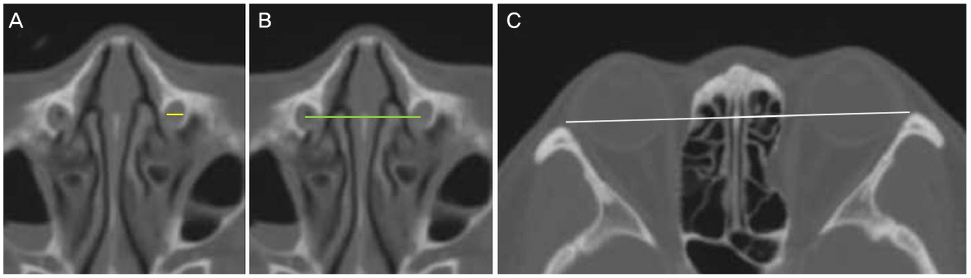
Figure 1 Bony nasolacrimal duct (BNLD) measurements with computed tomography (CT). (A) Axial CT image in bone window demonstrates BNLD transverse diameter (yellow line) measurement. (B) It reveals distance between the proximal BNLD (green line). (C) It shows inter-frontozygomatic suture distance (white line).

Figure 2 Bony nasolacrimal duct (BNLD) measurements with computed tomography (CT). (A) Sagittal CT image reveals BNLD length (orange line) and proximal, minimal, distal BNLD anteroposterior diameter (yellow lines) measurements, respectively. ‘a’ demonstrates measurement of sagittal orientation angle between long axis (white line) of BNLD and nasal floor (black line). (B) Coronal CT image shows relative lacrimal sac-BNLD orientation angle ‘b’ between long axis of lacrimal sac fossa (green line) and BNLD (white line). ‘c’ reveals angle between long axis of BNLD (white line) and nasal floor (black line). (C) It demonstrates angle (red line) between the bony inferior turbinate and upper part of the medial wall of the maxillary sinus.
Reference
-
1. Linberg JV, McCormick SA. Primary acquired nasolacrimal duct obstruction. A clinicopathologic report and biopsy technique. Ophthalmology. 1986; 93:1055–1063.2. Ohtomo K, Ueta T, Toyama T, Nagahara M. Predisposing factors for primary acquired nasolacrimal duct obstruction. Graefes Arch Clin Exp Ophthalmol. 2013; 251:1835–1839.3. Seider N, Miller B, Beiran I. Topical glaucoma therapy as a risk factor for nasolacrimal duct obstruction. Am J Ophthalmol. 2008; 145:120–123.4. Bulbul E, Yazici A, Yanik B, et al. Morphometric evaluation of bony nasolacrimal canal in a Caucasian population with primary acquired nasolacrimal duct obstruction: A Multidetector Computed Tomography Study. Korean J Radiol. 2016; 17:271–276.5. Gul A, Aslan K, Karli R, et al. A possible cause of nasolacrimal duct obstruction: narrow angle between inferior turbinate and upper part of the medial wall of the maxillary sinus. Curr Eye Res. 2016; 41:729–733.6. Janssen AG, Mansour K, Bos JJ, Castelijns JA. Diameter of the bony lacrimal canal: normal values and values related to nasolacrimal duct obstruction: assessment with CT. AJNR Am J Neuroradiol. 2001; 22:845–850.7. McCormick A, Sloan B. The diameter of the nasolacrimal canal measured by computed tomography: gender and racial differences. Clin Exp Ophthalmol. 2009; 37:357–361.8. Shigeta K, Takegoshi H, Kikuchi S. Sex and age differences in the bony nasolacrimal canal: an anatomical study. Arch Ophthalmol. 2007; 125:1677–1681.9. Estes JL, Tsiouris AJ, Christos PJ, Lelli GJ. Three-dimensional volumetric assessment of the nasolacrimal duct in patients with obstruction. Ophthal Plast Reconstr Surg. 2015; 31:211–214.10. George M, Phillips CI. Epiphora and the bony naso-lacrimal canal. Br J Ophthalmol. 1956; 40:673–680.11. Fasina O, Ogbole GI. CT assessment of the nasolacrimal canal in a black African Population. Ophthal Plast Reconstr Surg. 2013; 29:231–233.12. Groessl SA, Sires BS, Lemke BN. An anatomical basis for primary acquired nasolacrimal duct obstruction. Arch Ophthalmol. 1997; 115:71–74.13. Post RH. Tear duct size differences of age, sex and race. Am J Phys Anthropol. 1969; 30:85–88.14. Takahashi Y, Kakizaki H, Nakano T. Bony nasolacrimal duct entrance diameter: gender difference in cadaveric study. Ophthal Plast Reconstr Surg. 2011; 27:204–205.15. Takahashi Y, Nakamura Y, Nakano T, et al. The narrowest part of the bony nasolacrimal canal: an anatomical study. Ophthal Plast Reconstr Surg. 2013; 29:318–322.16. Ramey NA, Hoang JK, Richard MJ. Multidetector CT of nasolacrimal canal morphology: normal variation by age, gender, and race. Ophthal Plast Reconstr Surg. 2013; 29:475–480.17. Lee H, Ha S, Lee Y, et al. Anatomical and morphometric study of the bony nasolacrimal canal using computed tomography. Ophthalmologica. 2012; 227:153–159.18. Takahashi Y, Nakata K, Miyazaki H, et al. Comparison of bony nasolacrimal canal narrowing with or without primary acquired nasolacrimal duct obstruction in a Japanese population. Ophthal Plast Reconstr Surg. 2014; 30:434–438.19. Yong AM, Zhao DB, Siew SC, et al. Assessment of bony nasolacrimal parameters among Asians. Ophthal Plast Reconstr Surg. 2014; 30:322–327.
- Full Text Links
-
- Actions
-
Cited
- CITED
-
- Close
- Share
-
- Similar articles
-
- A Case of Nasolacrimal Duct Obstruction after Two-Jaw Surgery
- A Case of Angioleiomyoma in Nasolacrimal Duct
- The Methods of Insertiong the Root-type Lacrimal Tube and an Artificial nasolacrimal duct into the Men's
- A Case of Nasolacrimal Duct Obstruction Caused by a Lacrimal Sac Retention Cyst
- Nasolacrimal Duct Obstruction Following Midfacial Autologous Fat Injection
