Polyp Detection, Characterization, and Management Using Narrow-Band Imaging with/without Magnification
- Affiliations
-
- 1Gastrointestinal Center and Institution of Minimally Invasive Endoscopic Care (iMEC), Sano Hospital, Kobe, Japan. barce10_ronald@yahoo.co.jp
- KMID: 2380405
- DOI: http://doi.org/10.5946/ce.2015.48.6.491
Abstract
- Narrow-band imaging (NBI) is a new imaging technology that was developed in 2006 and has since spread worldwide. Because of its convenience, NBI has been replacing the role of chromoendoscopy. Here we review the efficacy of NBI with/without magnification for detection, characterization, and management of colorectal polyps, and future perspectives for the technology, including education. Recent studies have shown that the next-generation NBI system can detect significantly more colonic polyps than white light imaging, suggesting that NBI may become the modality of choice from the beginning of screening. The capillary pattern revealed by NBI, and the NBI International Colorectal Endoscopic classification are helpful for prediction of histology and for estimating the depth of invasion of colorectal cancer. However, NBI with magnifying colonoscopy is not superior to magnifying chromoendoscopy for estimation of invasion depth. Currently, therefore, chromoendoscopy should also be performed additionally if deep submucosal invasive cancer is suspected. If endoscopists become able to accurately estimate colorectal polyp pathology using NBI, this will allow adenomatous polyps to be resected and discarded; thus, reducing both the risk of polypectomy and costs. In order to achieve this goal, a suitable system for education and training in in vivo diagnostics will be necessary.
MeSH Terms
Figure
-
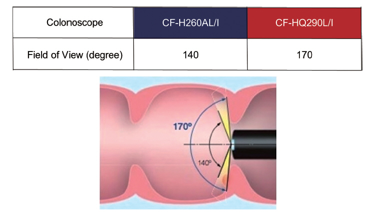
Fig. 1. Comparison of the field of view between the Olympus 260 and 290 series colonoscopes. The newly developed 290 series colonoscopes have a 170º field of view, 30º wider than the previous 260 series colonoscopes. This can help physicians to detect mucosal changes more rapidly, with less need for angulation.
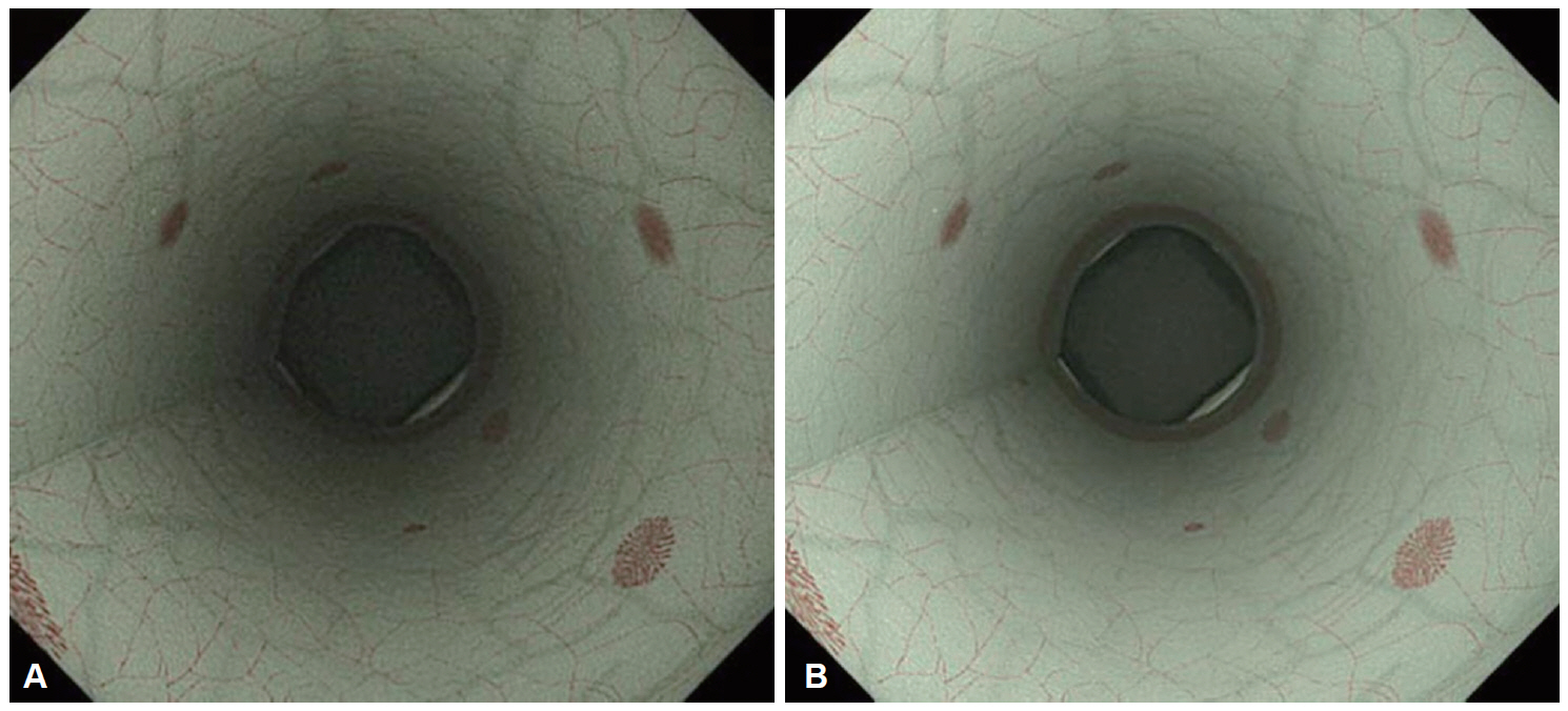
Fig. 2. Comparison of colonoscopic views using narrow-band imaging (NBI) between the Olympus 260 and 290 series endoscopy systems. (A) Colonoscopic view using NBI in the Olympus 260 series endoscopy system (EVIS LUCERA SPECTRUM). (B) Colonoscopic view using NBI in the Olympus 290 series endoscopy system (EVIS LUCERA ELITE). Colonoscopic views using NBI in the newly developed 290 series endoscopy system are brighter and have a higher resolution than those in the 260 series.
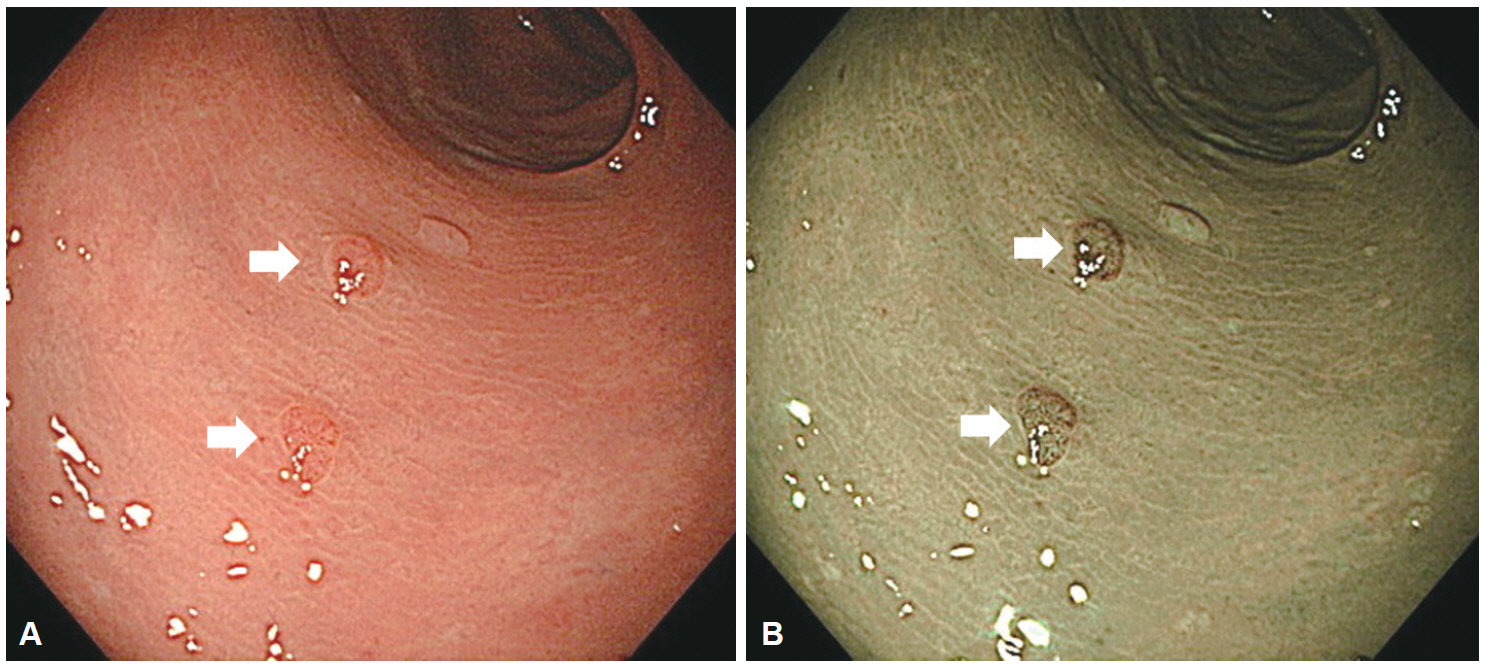
Fig. 3. Colonoscopic views using high-definition white light (HD-WL) and high-definition narrow-band imaging (HD-NBI) in the Olympus 290 series endoscopy system. (A) Colonoscopic view using HD-WL. (B) Colonoscopic view using HD-NBI. Colorectal neoplastic lesions are recognized as pale-reddish or reddish areas using WL, and as brownish areas using NBI, which is caused by mucosal microvascular dilatation and neoplastic angiogenesis. White arrows indicate neoplastic lesions.
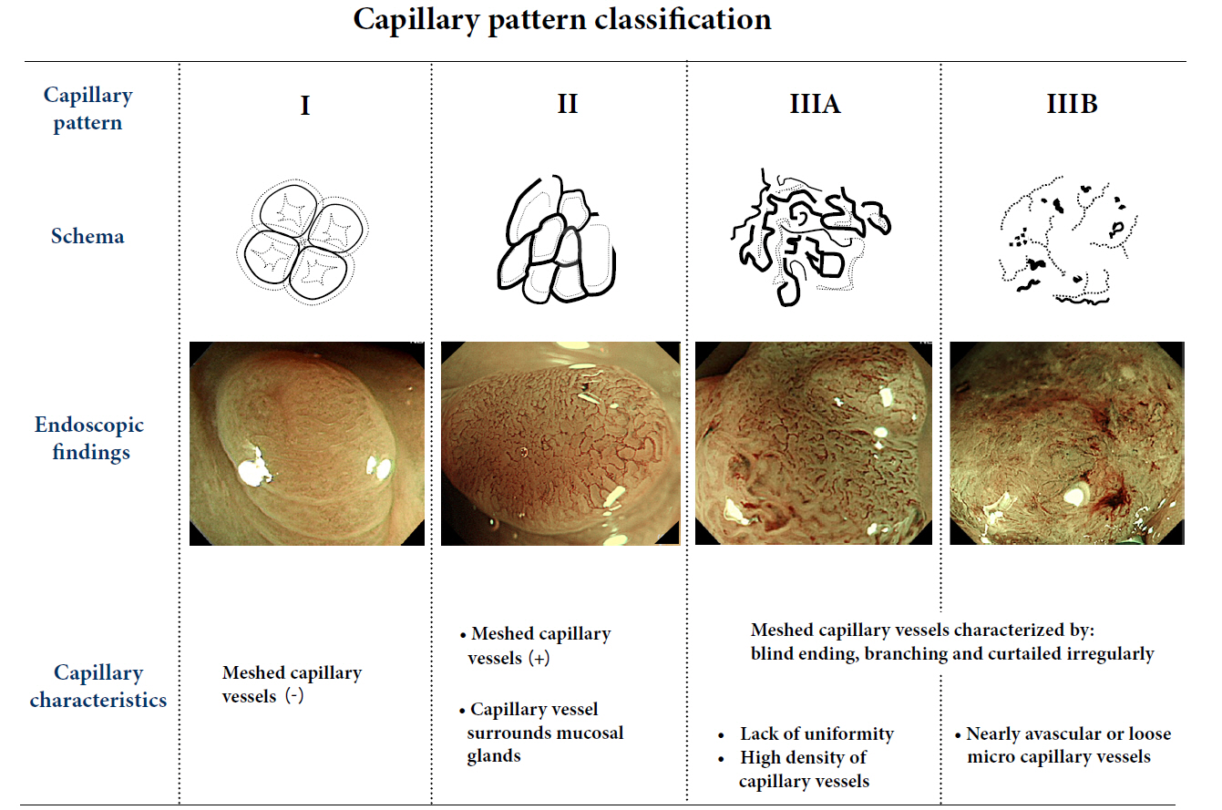
Fig. 4. Capillary pattern classification.
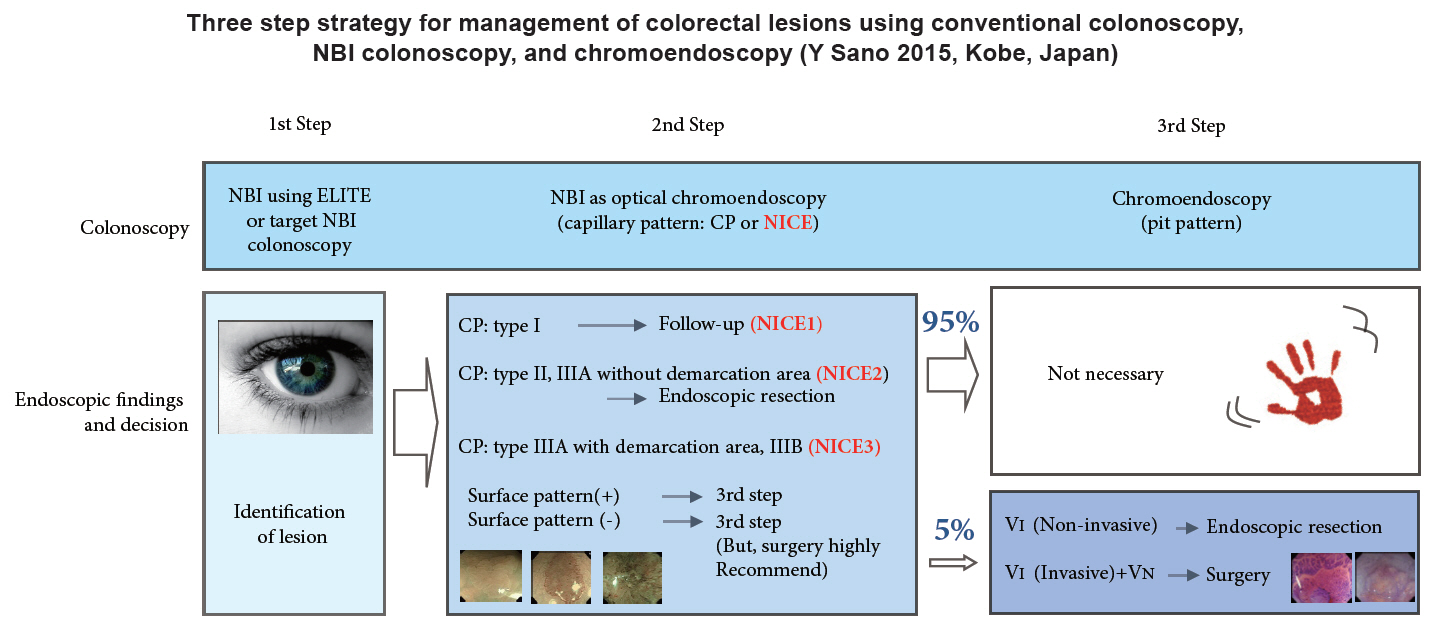
Fig. 5. Three-step strategy of narrow-band imaging (NBI). Adapted from Iwatate et al. [23] ELITE, Olympus 290 series endoscopy system EVIS LUCERA ELITE; NICE, NBI International Colorectal Endoscopic.
Reference
-
1. New diagnostic method based on color imaging using narrow band imaging (NBI) system for gastrointestinal tract. Gastrointest Endosc. 2001; 53:AB125.2. Masaki T, Katada C, Nakayama M, et al. Narrow band imaging in the diagnosis of intra-epithelial and invasive laryngeal squamous cell carcinoma: a preliminary report of two cases. Auris Nasus Larynx. 2009; 36:712–716.
Article3. Gono K, Obi T, Yamaguchi M, et al. Appearance of enhanced tissue features in narrow-band endoscopic imaging. J Biomed Opt. 2004; 9:568–577.
Article4. Folkman J. Tumor angiogenesis: therapeutic implications. N Engl J Med. 1971; 285:1182–1186.
Article5. Folkman J, Watson K, Ingber D, Hanahan D. Induction of angiogenesis during the transition from hyperplasia to neoplasia. Nature. 1989; 339:58–61.6. Aotake T, Lu CD, Chiba Y, Muraoka R, Tanigawa N. Changes of angiogenesis and tumor cell apoptosis during colorectal carcinogenesis. Clin Cancer Res. 1999; 5:135–142.7. Kudo S, Tamura S, Nakajima T, Yamano H, Kusaka H, Watanabe H. Diagnosis of colorectal tumorous lesions by magnifying endoscopy. Gastrointest Endosc. 1996; 44:8–14.
Article8. Pasha SF, Leighton JA, Das A, et al. Comparison of the yield and miss rate of narrow band imaging and white light endoscopy in patients undergoing screening or surveillance colonoscopy: a meta-analysis. Am J Gastroenterol. 2012; 107:363–370.
Article9. Nagorni A, Bjelakovic G, Petrovic B. Narrow band imaging versus conventional white light colonoscopy for the detection of colorectal polyps. Cochrane Database Syst Rev. 2012; 1:CD008361.
Article10. Horimatsu T, Sano Y, Tanaka S, et al. Next-generation narrow band imaging system for colonic polyp detection: a prospective multicenter randomized trial. Int J Colorectal Dis. 2015; 30:947–954.
Article11. Leung WK, Lo OS, Liu KS, et al. Detection of colorectal adenoma by narrow band imaging (HQ190) vs. high-definition white light colonoscopy: a randomized controlled trial. Am J Gastroenterol. 2014; 109:855–863.
Article12. Muto M, Minashi K, Yano T, et al. Early detection of superficial squamous cell carcinoma in the head and neck region and esophagus by narrow band imaging: a multicenter randomized controlled trial. J Clin Oncol. 2010; 28:1566–1572.
Article13. Fu KI, Sano Y, Kato S, et al. Chromoendoscopy using indigo carmine dye spraying with magnifying observation is the most reliable method for differential diagnosis between non-neoplastic and neoplastic colorectal lesions: a prospective study. Endoscopy. 2004; 36:1089–1093.
Article14. Sano Y, Emura F, Ikematsu H. Narrow band imaging. In : Waye JD, Rex DX, Williams CB, editors. Colonoscopy: Principles and Practice. Oxford: Blackwell;2009. p. 514–526.15. Sano Y, Ikematsu H, Fu KI, et al. Meshed capillary vessels by use of narrow-band imaging for differential diagnosis of small colorectal polyps. Gastrointest Endosc. 2009; 69:278–283.
Article16. Katagiri A, Fu KI, Sano Y, et al. Narrow band imaging with magnifying colonoscopy as diagnostic tool for predicting histology of early colorectal neoplasia. Aliment Pharmacol Ther. 2008; 27:1269–1274.
Article17. Ikematsu H, Matsuda T, Emura F, et al. Efficacy of capillary pattern type IIIA/IIIB by magnifying narrow band imaging for estimating depth of invasion of early colorectal neoplasms. BMC Gastroenterol. 2010; 10:33.
Article18. Matsuda T, Fujii T, Saito Y, et al. Efficacy of the invasive/non-invasive pattern by magnifying chromoendoscopy to estimate the depth of invasion of early colorectal neoplasms. Am J Gastroenterol. 2008; 103:2700–2706.
Article19. Rex DK. Narrow-band imaging without optical magnification for histologic analysis of colorectal polyps. Gastroenterology. 2009; 136:1174–1181.
Article20. Ignjatovic A, East JE, Suzuki N, Vance M, Guenther T, Saunders BP. Optical diagnosis of small colorectal polyps at routine colonoscopy (Detect InSpect ChAracterise Resect and Discard; DISCARD trial): a prospective cohort study. Lancet Oncol. 2009; 10:1171–1178.
Article21. Hewett DG, Kaltenbach T, Sano Y, et al. Validation of a simple classification system for endoscopic diagnosis of small colorectal polyps using narrow-band imaging. Gastroenterology. 2012; 143:599–607.e1.
Article22. Hayashi N, Tanaka S, Hewett DG, et al. Endoscopic prediction of deep submucosal invasive carcinoma: validation of the narrow-band imaging international colorectal endoscopic (NICE) classification. Gastrointest Endosc. 2013; 78:625–632.
Article23. Iwatate M, Ikumoto T, Hattori S, Sano W, Sano Y, Fujimori T. NBI and NBI combined with magnifying colonoscopy. Diagn Ther Endosc. 2012; 2012:173269.
Article24. Higashi R, Uraoka T, Kato J, et al. Diagnostic accuracy of narrow-band imaging and pit pattern analysis significantly improved for less-experienced endoscopists after an expanded training program. Gastrointest Endosc. 2010; 72:127–135.
Article25. Raghavendra M, Hewett DG, Rex DK. Differentiating adenomas from hyperplastic colorectal polyps: narrow-band imaging can be learned in 20 minutes. Gastrointest Endosc. 2010; 72:572–576.
Article26. Ignjatovic A, Thomas-Gibson S, East JE, et al. Development and validation of a training module on the use of narrow-band imaging in differentiation of small adenomas from hyperplastic colorectal polyps. Gastrointest Endosc. 2011; 73:128–133.
Article27. Rastogi A, Rao DS, Gupta N, et al. Impact of a computer-based teaching module on characterization of diminutive colon polyps by using narrow-band imaging by non-experts in academic and community practice: a video-based study. Gastrointest Endosc. 2014; 79:390–398.28. Ladabaum U, Fioritto A, Mitani A, et al. Real-time optical biopsy of colon polyps with narrow band imaging in community practice does not yet meet key thresholds for clinical decisions. Gastroenterology. 2013; 144:81–91.
Article29. Kuiper T, Marsman WA, Jansen JM, et al. Accuracy for optical diagnosis of small colorectal polyps in nonacademic settings. Clin Gastroenterol Hepatol. 2012; 10:1016–1020.
Article30. Dai J, Shen YF, Sano Y, et al. Evaluation of narrow-band imaging in the diagnosis of colorectal lesions: is a learning curve involved? Dig Endosc. 2013; 25:180–188.
Article31. Machida H, Sano Y, Hamamoto Y, et al. Narrow-band imaging in the diagnosis of colorectal mucosal lesions: a pilot study. Endoscopy. 2004; 36:1094–1098.
Article32. Su MY, Hsu CM, Ho YP, Chen PC, Lin CJ, Chiu CT. Comparative study of conventional colonoscopy, chromoendoscopy, and narrow-band imaging systems in differential diagnosis of neoplastic and nonneoplastic colonic polyps. Am J Gastroenterol. 2006; 101:2711–2716.
Article33. Apel D, Jakobs R, Schilling D, et al. Accuracy of high-resolution chromoendoscopy in prediction of histologic findings in diminutive lesions of the rectosigmoid. Gastrointest Endosc. 2006; 63:824–828.
Article34. Tischendorf JJ, Wasmuth HE, Koch A, Hecker H, Trautwein C, Winograd R. Value of magnifying chromoendoscopy and narrow band imaging (NBI) in classifying colorectal polyps: a prospective controlled study. Endoscopy. 2007; 39:1092–1096.
Article35. De Palma GD, Rega M, Masone S, et al. Conventional colonoscopy and magnified chromoendoscopy for the endoscopic histological prediction of diminutive colorectal polyps: a single operator study. World J Gastroenterol. 2006; 12:2402–2405.36. Rex DK, Kahi C, O’Brien M, et al. The American Society for Gastrointestinal Endoscopy PIVI (Preservation and Incorporation of Valuable Endoscopic Innovations) on real-time endoscopic assessment of the histology of diminutive colorectal polyps. Gastrointest Endosc. 2011; 73:419–422.
Article37. ASGE Technology Committee, Abu Dayyeh BK, Thosani N, et al. ASGE Technology Committee systematic review and meta-analysis assessing the ASGE PIVI thresholds for adopting real-time endoscopic assessment of the histology of diminutive colorectal polyps. Gastrointest Endosc. 2015; 81:502.e1–502.e16.
Article38. Iwatate M, Sano Y, Hattori S, et al. The addition of high magnifying endoscopy improves rates of high confidence optical diagnosis of colorectal polyps. Endosc Int Open. 2015; 3:E140–E145.
Article
- Full Text Links
-
- Actions
-
Cited
- CITED
-
- Close
- Share
-
- Similar articles
-
- Advanced Imaging Technology Other than Narrow Band Imaging
- Usage of Narrow Band Imaging System in the Colorectum
- Narrow Band Imaging and White Light Colonoscopy for Detection of Polyps
- Differential Diagnosis of Colorectal Polyps with Narrow Band Imaging Colonoscopy without Magnification
- Usefulness of Narrow-Band Imaging in Endoscopic Submucosal Dissection of the Stomach
