Maxillomandibular arch width differences at estimated centers of resistance: Comparison between normal occlusion and skeletal Class III malocclusion
- Affiliations
-
- 1Department of Orthodontics, College of Dentistry, Yonsei University, Seoul, Korea.
- 2Department of Orthodontics, Institute of Craniofacial Deformity, College of Dentistry, Yonsei University, Seoul, Korea.
- 3Department of Orthodontics, University of Aarhus, Aarhus, Denmark.
- KMID: 2379650
- DOI: http://doi.org/10.4041/kjod.2017.47.3.167
Abstract
OBJECTIVE
To evaluate the differences in maxillomandibular transverse measurements at either the crown or the estimated center of resistance (CR), and to compare values between normal occlusion and Class III malocclusion groups.
METHODS
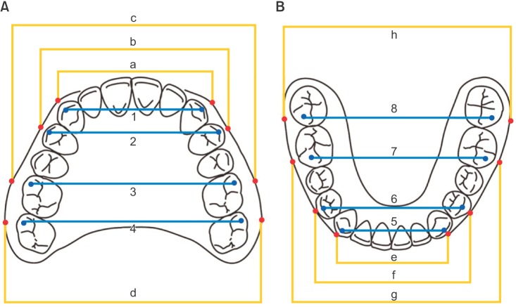
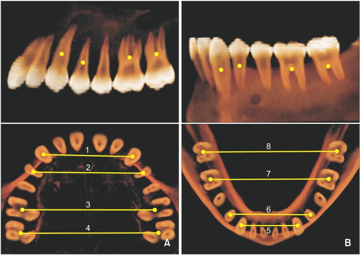
Dental casts and computed tomography (CT) data from 30 individuals with normal occlusion and 30 with skeletal Class III malocclusions were evaluated. Using the casts, dental arch widths (DAWs) were measured from the cusp tips, and basal arch widths (BAWs-cast) were measured as the distance between the points at the mucogingival junction adjacent to the respective cusp tips. The BAWs determined from CT (BAWs-CT) images were measured from the estimated CRs of the teeth.
RESULTS
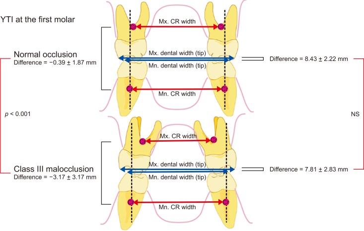
None of the DAW measurements or maxillomandibular DAW differences showed statistically significant intergroup differences. In contrast, the maxillary BAWs-CT and BAWs-cast were lesser in the Class III malocclusion group than in the normal occlusion group. The mandibular BAWs-CT were significantly greater in the Class III malocclusion group than in the normal occlusion group. Moreover, the maxillomandibular BAW differences on both CT and cast showed significant intergroup differences in all transverse measurements.
CONCLUSIONS
The maxillomandibular DAW differences showed no significant intergroup differences. In contrast, the maxillomandibular BAW differences on both CT and cast showed significant intergroup differences in all transverse measurements. The maxillomandibular BAW differences at the estimated CRs, measured using CT or casts, can reveal underlying transverse maxillary basal arch deficiencies in patients with skeletal Class III malocclusions.
Keyword
MeSH Terms
Figure
Cited by 4 articles
-
Stability of bimaxillary surgery involving intraoral vertical ramus osteotomy with or without presurgical miniscrew-assisted rapid palatal expansion in adult patients with skeletal Class III malocclusion
Yoon-Soo Ahn, Sung-Hwan Choi, Kee-Joon Lee, Young-Soo Jung, Hyoung-Seon Baik, Hyung-Seog Yu
Korean J Orthod. 2020;50(5):304-313. doi: 10.4041/kjod.2020.50.5.304.Effects of bodily retraction of mandibular incisors versus mandibular setback surgery on pharyngeal airway space: A comparative study
Byeong-Tak Keum, Sung-Hwan Choi, Yoon Jeong Choi, Hyoung-Seon Baik, Kee-Joon Lee
Korean J Orthod. 2017;47(6):344-352. doi: 10.4041/kjod.2017.47.6.344.Evaluation of the effects of miniscrew incorporation in palatal expanders for young adults using finite element analysis
Eui-Hyang Seong, Sung-Hwan Choi, Hee-Jin Kim, Hyung-Seog Yu, Young-Chel Park, Kee-Joon Lee
Korean J Orthod. 2018;48(2):81-89. doi: 10.4041/kjod.2018.48.2.81.Nonsurgical maxillary expansion in a 60-year-old patient with gingival recession and crowding
Harim Kim, Sun-Hyung Park, Jae Hyun Park, Kee-Joon Lee
Korean J Orthod. 2021;51(3):217-227. doi: 10.4041/kjod.2021.51.3.217.
Reference
-
1. Franchi L, Baccetti T. Transverse maxillary deficiency in Class II and Class III malocclusions: a cephalometric and morphometric study on postero-anterior films. Orthod Craniofac Res. 2005; 8:21–28. PMID: 15667642.
Article2. Proffit WR, Phillips C, Prewitt JW, Turvey TA. Stability after surgical-orthodontic correction of skeletal Class III malocclusion. 2. Maxillary advancement. Int J Adult Orthodon Orthognath Surg. 1991; 6:71–80. PMID: 1811032.3. McNamara JA. Maxillary transverse deficiency. Am J Orthod Dentofacial Orthop. 2000; 117:567–570. PMID: 10799117.4. Uysal T, Usumez S, Memili B, Sari Z. Dental and alveolar arch widths in normal occlusion and Class III malocclusion. Angle Orthod. 2005; 75:809–813. PMID: 16279827.5. Ricketts RM. Perspectives in the clinical application of cephalometrics. The first fifty years. Angle Orthod. 1981; 51:115–150. PMID: 6942666.6. Hesby RM, Marshall SD, Dawson DV, Southard KA, Casko JS, Franciscus RG, et al. Transverse skeletal and dentoalveolar changes during growth. Am J Orthod Dentofacial Orthop. 2006; 130:721–731. PMID: 17169734.
Article7. Leonardi R, Annunziata A, Caltabiano M. Landmark identification error in posteroanterior cephalometric radiography. A systematic review. Angle Orthod. 2008; 78:761–765. PMID: 18302479.8. Jung PK, Lee GC, Moon CH. Comparison of cone-beam computed tomography cephalometric measurements using a midsagittal projection and conventional two-dimensional cephalometric measurements. Korean J Orthod. 2015; 45:282–288. PMID: 26629474.
Article9. Kusayama M, Motohashi N, Kuroda T. Relationship between transverse dental anomalies and skeletal asymmetry. Am J Orthod Dentofacial Orthop. 2003; 123:329–337. PMID: 12637905.
Article10. Tyan S, Park HS, Janchivdorj M, Han SH, Kim SJ, Ahn HW. Three-dimensional analysis of molar compensation in patients with facial asymmetry and mandibular prognathism. Angle Orthod. 2016; 86:421–430. PMID: 26192894.
Article11. Burstone CR. Deep overbite correction by intrusion. Am J Orthod. 1977; 72:1–22. PMID: 267433.
Article12. Melsen B, Agerbaek N, Markenstam G. Intrusion of incisors in adult patients with marginal bone loss. Am J Orthod Dentofacial Orthop. 1989; 96:232–241. PMID: 2773869.
Article13. Smith RJ, Burstone CJ. Mechanics of tooth movement. Am J Orthod. 1984; 85:294–307. PMID: 6585147.
Article14. Jo AR, Mo SS, Lee KJ, Sung SJ, Chun YS. Finite-element analysis of the center of resistance of the mandibular dentition. Korean J Orthod. 2017; 47:21–30. PMID: 28127536.
Article15. van Steenbergen E, Burstone CJ, Prahl-Andersen B, Aartman IH. Influence of buccal segment size on prevention of side effects from incisor intrusion. Am J Orthod Dentofacial Orthop. 2006; 129:658–665. PMID: 16679206.
Article16. Lee KJ, Joo E, Kim KD, Lee JS, Park YC, Yu HS. Computed tomographic analysis of tooth-bearing alveolar bone for orthodontic miniscrew placement. Am J Orthod Dentofacial Orthop. 2009; 135:486–494. PMID: 19361735.
Article17. Vanarsdall RL Jr. Transverse dimension and long-term stability. Semin Orthod. 1999; 5:171–180. PMID: 10860069.
Article18. Burstone CJ, Choy K. The biomechanical foundation of clinical orthodontics. Chicago: Quintessence Publishing Co, Inc.;2015.19. Lim MY, Lim SH. Comparison of model analysis measurements among plaster model, laser scan digital model, and cone beam CT image. Korean J Orthod. 2009; 39:6–17.
Article20. Barrett JF, Keat N. Artifacts in CT: recognition and avoidance. Radiographics. 2004; 24:1679–1691. PMID: 15537976.
Article21. Kuntz TR, Staley RN, Bigelow HF, Kremenak CR, Kohout FJ, Jakobsen JR. Arch widths in adults with Class I crowded and Class III malocclusions compared with normal occlusions. Angle Orthod. 2008; 78:597–603. PMID: 18302456.
Article22. Lee HK, Son WS. A study on basal and dental arch width in skeletal class III malocclusion. Korean J Orthod. 2002; 32:117–127.23. Masumoto T, Hayashi I, Kawamura A, Tanaka K, Kasai K. Relationships among facial type, buccolingual molar inclination, and cortical bone thickness of the mandible. Eur J Orthod. 2001; 23:15–23. PMID: 11296507.
Article24. Suk KE, Park JH, Bayome M, Nam YO, Sameshima GT, Kook YA. Comparison between dental and basal arch forms in normal occlusion and Class III malocclusions utilizing cone-beam computed tomography. Korean J Orthod. 2013; 43:15–22. PMID: 23504406.
Article25. Robert WE. Bone physiology, metabolism, biomechanics in orthodontic practice. In : Graber LE, Vanarsdall RL, Vig KW, editors. Orthodontics: current principles and techniques. 5th ed. Philadelphia, PA: Elsevier/Mosby;2012.26. Proffit WR. Equilibrium theory revisited: factors influencing position of the teeth. Angle Orthod. 1978; 48:175–186. PMID: 280125.27. Ovsenik M, Primožič J. How to push the limits in the transverse dimension? Facial asymmetry, palatal volume and tongue posture in children with unilateral posterior cross bite: a three-dimensional evaluation of early treatment. Orthod Fr. 2014; 85:139–149. PMID: 24923214.28. Braun S, Hnat WP, Fender DE, Legan HL. The form of the human dental arch. Angle Orthod. 1998; 68:29–36. PMID: 9503132.
- Full Text Links
- Actions
-
Cited
- CITED
-
- Close
- Share
- Similar articles
-
- Study of horizontal skeletal pattern and dental arch in skeletal Class III malocclusion patients
- A Study on Basal and Dental Arch Width in Skeletal Class III Malocclusion
- Posteroanterior cephalometric characteristics in skeletal Class III malocclusion
- A study on the difference of the skeletal maturity in normal occlusion and malocclusion
- A study on the maxillary dental arch and palatal vault with malocculsions


