Improved Software to Browse the Serial Medical Images for Learning
- Affiliations
-
- 1Department of Computer Engineering, Inha University, Incheon, Korea.
- 2Department of Anatomy, Ajou University School of Medicine, Suwon, Korea. bschung@ajou.ac.kr
- 3Department of Anatomy, Dongguk University School of Medicine, Gyeongju, Korea.
- KMID: 2379617
- DOI: http://doi.org/10.3346/jkms.2017.32.7.1195
Abstract
- The thousands of serial images used for medical pedagogy cannot be included in a printed book; they also cannot be efficiently handled by ordinary image viewer software. The purpose of this study was to provide browsing software to grasp serial medical images efficiently. The primary function of the newly programmed software was to select images using 3 types of interfaces: buttons or a horizontal scroll bar, a vertical scroll bar, and a checkbox. The secondary function was to show the names of the structures that had been outlined on the images. To confirm the functions of the software, 3 different types of image data of cadavers (sectioned and outlined images, volume models of the stomach, and photos of the dissected knees) were inputted. The browsing software was downloadable for free from the homepage (anatomy.co.kr) and available off-line. The data sets provided could be replaced by any developers for their educational achievements. We anticipate that the software will contribute to medical education by allowing users to browse a variety of images.
Keyword
MeSH Terms
Figure
-
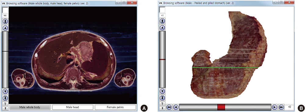
Fig. 1 Initial views of the 2 new browsing software packages. (A) The software shows horizontal sectioned image with the x-axis represented by the 3 buttons. The image file shown in the initial view is named “1_851.png.” The “1” indicates the first button (male whole body), and “851” indicates the 851st sectioned image. (B) The software shows stomach volume model with the x-axis as the scroll bar. The image file shown in the initial view is named “0_0.png,” where the first “0” indicates 0 degrees of rotation and the second “0” indicates the volume model with no peeling or piling.
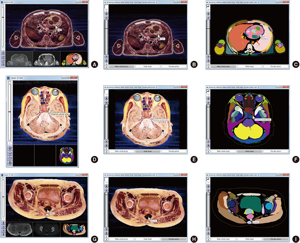
Fig. 2 Comparison of previous and new software that automatically label the names of the outlined structures of 3 cadavers. (A, B, C) Male whole body. (D, E, F) Male head. (G, H, I) Female pelvis. (A, D, G) Previous software to simultaneously show the sectioned and color-filled images. (B, E, H) New software to show the sectioned images when the checkbox is off. (C, F, I) New software to show the color-filled images when the checkbox is on.
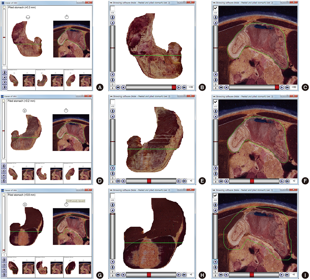
Fig. 3 Comparison of previous and new software that show the volume models and sectioned images of stomach. (A, B, C) Posterior view of stomach. (D, E, F) Anterior view of stomach. (G, H, I) Anterior view of piled stomach. (A, D, G) Previous software to simultaneously show the volume models and the sectioned image of the stomach. (B, E, H) New software to alternatively show the volume models when the checkbox is off. (C, F, I) New software to alternatively show the sectioned image when the checkbox is on.
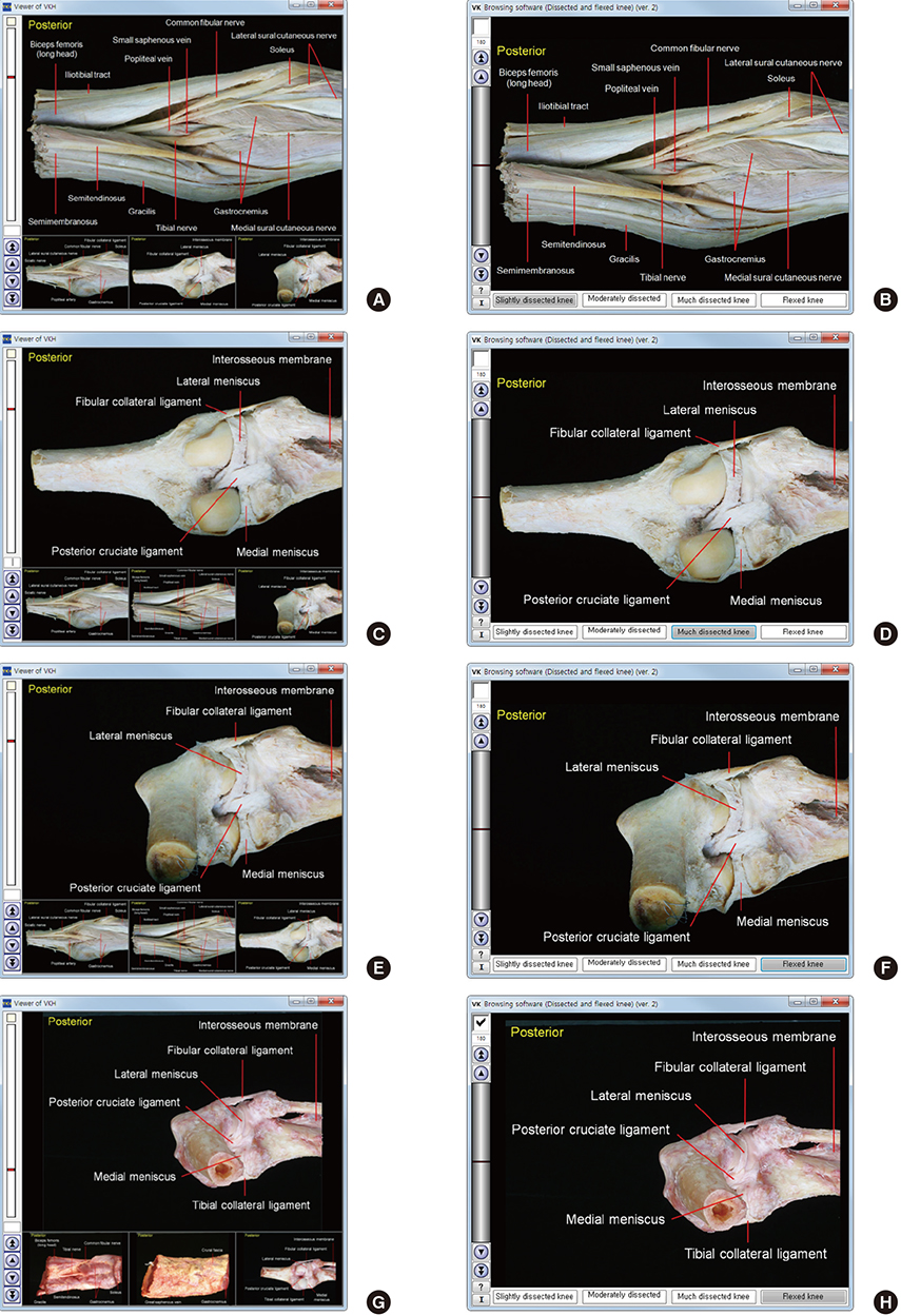
Fig. 4 Comparison of previous and new software that show cadaveric knees. (A, B) Slightly dissected knee of embalmed cadaver. (C, D) Much dissected knee of embalmed cadaver. (E, F) Flexed knee of embalmed cadaver. (G, H) Flexed knee of fresh cadaver. (A, C, E, G) Previous software. (B, D, F, H) New software.
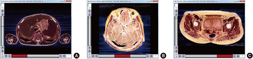
Fig. 5 Experimental software to show the sectioned images of 3 cadavers with the x-axis scroll bar. (A) Male whole body. (B) Male head. (C) Female pelvis.
Cited by 8 articles
-
Dawn of the Visible Monkey: Segmentation of the Rhesus Monkey for 2D and 3D Applications
Chung Yoh Kim, Ae-Kyoung Lee, Hyung-Do Choi, Jin Seo Park
J Korean Med Sci. 2020;35(15):e100. doi: 10.3346/jkms.2020.35.e100.Adaptations in Anatomy Education during COVID-19
Hyeijung Yoo, Dasom Kim, Young-Mee Lee, Im Joo Rhyu
J Korean Med Sci. 2020;36(1):e13. doi: 10.3346/jkms.2021.36.e13.Three Software Tools for Viewing Sectional Planes, Volume Models, and Surface Models of a Cadaver Hand
Beom Sun Chung, Min Suk Chung, Byeong-Seok Shin, Koojoo Kwon
J Korean Med Sci. 2018;33(8):. doi: 10.3346/jkms.2018.33.e64.Advanced Sectioned Images of a Cadaver Head with Voxel Size of 0.04 mm
Beom Sun Chung, Miran Han, Donghwan Har, Jin Seo Park
J Korean Med Sci. 2019;34(34):. doi: 10.3346/jkms.2019.34.e218.Real-Color Volume Models Made from Real-Color Sectioned Images of Visible Korean
Beom Sun Chung, Jin Seo Park
J Korean Med Sci. 2019;34(10):. doi: 10.3346/jkms.2019.34.e86.New Viewpoint of Surface Anatomy Using the Curved Sectional Planes of a Male Cadaver
Koojoo Kwon, Byeong-Seok Shin, Min Suk Chung, Beom Sun Chung
J Korean Med Sci. 2019;34(3):. doi: 10.3346/jkms.2019.34.e15.Effects of Reading a Free Electronic Book on Regional Anatomy with Schematics and Mnemonics on Student Learning
Beom Sun Chung, Ki Seok Koh, Chang-Seok Oh, Jin Seo Park, Jae-Ho Lee, Min Suk Chung
J Korean Med Sci. 2020;35(6):. doi: 10.3346/jkms.2020.35.e42.Homepage to distribute the anatomy learning contents including Visible Korean products, comics, and books
Beom Sun Chung, Min Suk Chung
Anat Cell Biol. 2018;51(1):7-13. doi: 10.5115/acb.2018.51.1.7.
Reference
-
1. Plaisant C, Carr D, Shneiderman B. Image-browser taxonomy and guidelines for designers. IEEE Softw. 1995; 12:21–32.2. Rorden C, Brett M. Stereotaxic display of brain lesions. Behav Neurol. 2000; 12:191–200.3. Mojsilovic A, Gomes J. Semantic based categorization, browsing and retrieval in medical image databases. Proc Int Conf Image Proc. 2002; 3:III145–8.4. Shin DS, Chung MS, Park HS, Park JS, Hwang SB. Browsing software of the visible Korean data used for teaching sectional anatomy. Anat Sci Educ. 2011; 4:327–332.5. Shin DS, Jang HG, Park JS, Park HS, Lee S, Chung MS. Accessible and informative sectioned images and surface models of a cadaver head. J Craniofac Surg. 2012; 23:1176–1180.6. Shin DS, Jang HG, Hwang SB, Har DH, Moon YL, Chung MS. Two-dimensional sectioned images and three-dimensional surface models for learning the anatomy of the female pelvis. Anat Sci Educ. 2013; 6:316–323.7. Chung BS, Park JS, Jang HG, Chung MS. Software to browse the pictures of two knees in diverse states of dissection, flexion and rotation. Int J Morphol. 2015; 33:1009–1015.8. Kwon K, Shin DS, Shin BS, Park HS, Lee S, Jang HG, Park JS, Chung MS. Virtual endoscopic and laparoscopic exploration of stomach wall based on a cadaver’s sectioned images. J Korean Med Sci. 2015; 30:658–661.9. Chung BS, Chung MS, Shin BS, Kwon K. Peeled and piled volume models of the kidney that show actual morphology. J Korean Med Sci. 2016; 31:1514–1515.10. Park JS, Chung MS, Hwang SB, Lee YS, Har DH, Park HS. Visible Korean human: improved serially sectioned images of the entire body. IEEE Trans Med Imaging. 2005; 24:352–360.11. Park JS, Chung MS, Shin DS, Har DH, Cho ZH, Kim YB, Han JY, Chi JG. Sectioned images of the cadaver head including the brain and correspondences with ultrahigh field 7.0 T MRIs. Proc IEEE Inst Electr Electron Eng. 2009; 97:1988–1996.12. Hwang SB, Chung MS, Hwang YI, Park HS, Har DH, Shin DS, Shin BS, Park JS. Improved sectioned images of the female pelvis showing detailed urogenital and neighboring structures. Korean J Phys Anthropol. 2010; 23:187–198.13. Chung BS, Kwon K, Shin BS, Chung MS. Peeled and piled volume models of the stomach made from a cadaver ‘s sectioned images. Int J Morphol. 2016; 34:939–944.14. Alvarez A, Gold GE, Tobin B, Desser TS. Software tools for interactive instruction in radiologic anatomy. Acad Radiol. 2006; 13:512–517.15. Biasutto SN, Caussa LI, Criado del Río LE. Teaching anatomy: cadavers vs. computers? Ann Anat. 2006; 188:187–190.
- Full Text Links
-
- Actions
-
Cited
- CITED
-
- Close
- Share
-
- Similar articles
-
- Three Software Tools for Viewing Sectional Planes, Volume Models, and Surface Models of a Cadaver Hand
- Serial Slice Images and Segmented Images of the Brainstem for Recognizing the Stereoscopic Morphology of its Nuclei and Tracts
- Three Dimensional MRI and Software for Studying Normal Anatomical Structures of an Entire Body
- Serially Sectioned and Segmented Images of the Mouse for Learning Mouse Anatomy
- Serially Sectioned Images of the Whole Body - Sixth Report: Browsing Software of the Serially Sectioned Images for Learning Sectional Anatomy
