Transarterial Embolization Treatment for Aberrant Systemic Arterial Supply to the Normal Lung: A Case Report and Literature Review
- Affiliations
-
- 1Department of Radiology, Dong-A University Hospital, Dong-A University College of Medicine, Busan, Korea. bhpark@dau.ac.kr
- KMID: 2379321
- DOI: http://doi.org/10.3348/jksr.2017.76.6.395
Abstract
- A 24-year-old man presented with dyspnea on exertion and intermittent blood-tinged sputum. He was diagnosed with aberrant systemic arterial supply to the normal lung (ASANL) based on the results of imaging studies. The patient was successfully treated with transarterial embolization using coils and a vascular plug and his symptoms disappeared during the follow-up. Herein, we reported the imaging findings of ASANL, differential diagnoses, and its treatment options. In addition, we reviewed the relevant literature.
MeSH Terms
Figure
-
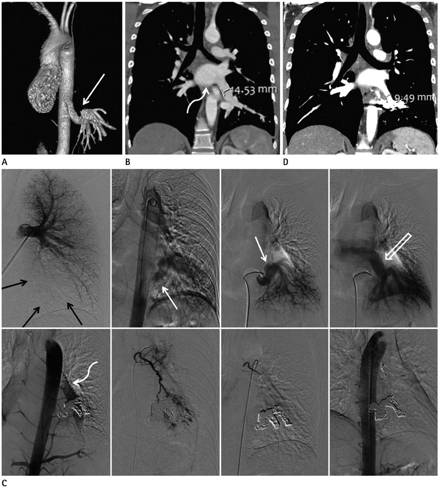
Fig. 1 Aberrant systemic arterial supply to the normal lung treated with transarterial embolization in a 24-year-old man. A, B. Initial chest CT. Volume rendering image (A) shows a 14-mm-sized, large tortuous vessel (arrow) arising from the descending thoracic aorta and supplying the basal segments of the left lower lobe. On lung window setting image, thickening of the interstitial markings with ground glass opacities, consistent with pulmonary congestion, was noted in the left lower lobe; courses of the bronchial tree in both lungs were normal (not shown). On coronal reformatted image (B), the left lower lobar pulmonary vein is dilated, as compared to the right lower lobar pulmonary vein (curved arrow), and normally drains to the left atrium. C. Images of conventional angiography (upper panel) and transarterial embolization (lower panel). Left pulmonary angiography shows hypoplasia or absence of the left lower pulmonary artery and decreased perfusion in the left lower lung zone (black arrows). Initial thoracic aortography and selective angiography of the aberrant systemic artery reveal an aberrant systemic artery (white arrows) arising from the descending thoracic aorta and supplying the left lower lung zone. On delayed phase, the enlarged left lower pulmonary vein (open arrow) that drains to the left atrium, is observed. On post-embolization aortography, the aberrant systemic artery is no longer visualized, and hypertrophied left bronchial artery (curved arrow) with arteriovenous shunt in the left lower lung zone, which was not revealed on the initial thoracic aortography, is observed. The left bronchial artery was selected, and embolization was conducted using microcoils and gelfoam particles. On final post-embolization aortography, the aberrant systemic artery and the hypertrophied bronchial artery are obliterated. D. Follow-up chest CT image at 4 months after treatment. On the coronal reformatted image after treatment, the left lower lobar pulmonary vein is decreased in diameter.
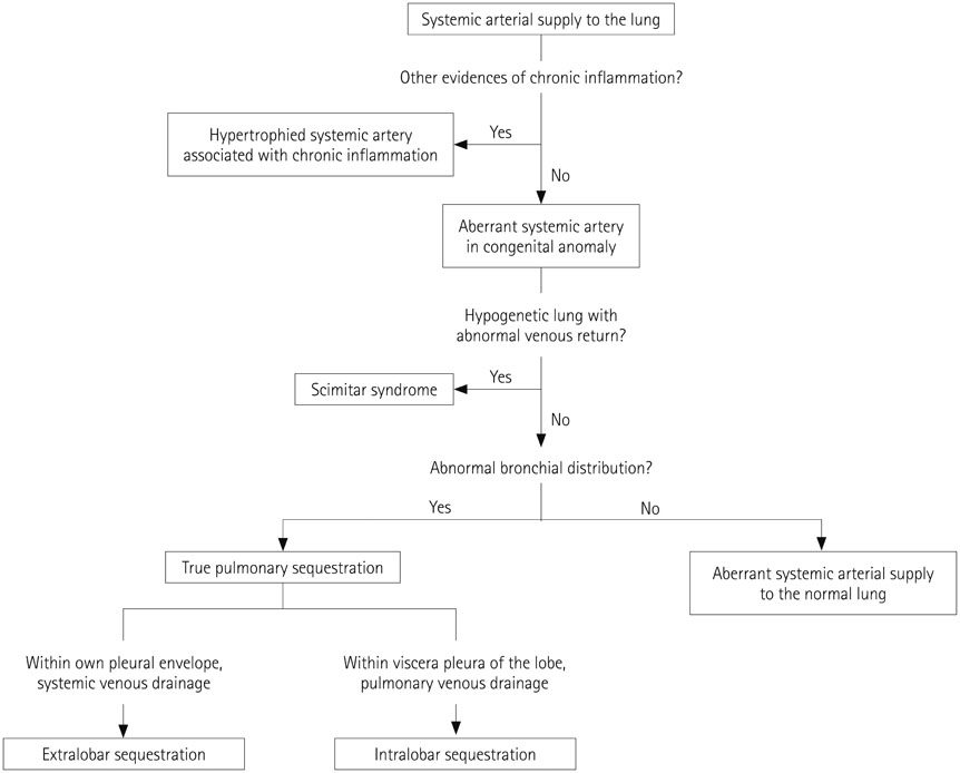
Fig. 2 A simplified algorithm for differential diagnosis of systemic arterial supply to the lung. The presence or absence of ancillary findings of chronic inflammation, abnormal venous return, and courses of the bronchial tree should be checked for differential diagnosis.
Reference
-
1. Kim JH, Kim SS, Ha KS, Bae J, Park Y. Anomalous arterial supply to normal basal segment of the right lower lobe: endovascular treatment with the amplatzer vascular plug. Tuberc Respir Dis (Seoul). 2014; 76:295–298.2. Muñoz JJ, García JA, Bentabol M, Padín MI, Serrano F. Endovascular treatment of hemoptysis by abnormal systemic pulmonary artery supply. Cardiovasc Intervent Radiol. 2008; 31:427–430.3. Brühlmann W, Weishaupt D, Goebel N, Imhof E. Therapeutic embolization of a systemic arterialization of lung without sequestration. Eur Radiol. 1998; 8:355–358.4. Jariwala P, Ramesh G, Sarat Chandra K. Congenital anomalous/aberrant systemic artery to pulmonary venous fistula: closure with vascular plugs & coil embolization. Indian Heart J. 2014; 66:95–103.5. Abe T, Mori K, Shiigai M, Okura N, Okamoto Y, Saida T, et al. Systemic arterial supply to the normal basal segments of the left lower lobe of the lung--treatment by coil embolization--and a literature review. Cardiovasc Intervent Radiol. 2011; 34:Suppl 2. S117–S121.6. Do KH, Goo JM, Im JG, Kim KW, Chung JW, Park JH. Systemic arterial supply to the lungs in adults: spiral CT findings. Radiographics. 2001; 21:387–402.7. Saida T, Ninomiya H, Hojo F, Nakayama M, Yamauchi T, Saida Y. Systemic arterial supply to the normal basal segments of the left lower lobe treated by coil embolization, with long-term follow-up. Radiat Med. 2006; 24:365–368.8. Pollak JS, Saluja S, Thabet A, Henderson KJ, Denbow N, White RI Jr. Clinical and anatomic outcomes after embolotherapy of pulmonary arteriovenous malformations. J Vasc Interv Radiol. 2006; 17:35–44. quiz 45.9. Brillet PY, Dumont P, Bouaziz N, Duhamel A, Laurent F, Remy J, et al. Pulmonary arteriovenous malformation treated with embolotherapy: systemic collateral supply at multidetector CT angiography after 2-20-year follow-up. Radiology. 2007; 242:267–276.10. Anil G, Taneja M, Tan AG. Endovascular treatment of isolated systemic arterial supply to normal lung with coil and glue embolisation. Br J Radiol. 2012; 85:e83–e86.
- Full Text Links
-
- Actions
-
Cited
- CITED
-
- Close
- Share
-
- Similar articles
-
- Treatment of Systemic Arterial Supply to Lower Lobe of Left Lung (Operation vs. Embolotherapy): Comparison of Two Cases and Literature Review
- A Case of Anomalous Systemic Arterial Supply to Normal Basal Segments of Left Lower Lobe
- A Case of Systemic Arterialization of the Lung without Sequestration
- One Case of Sustemic Arterialization of Lung Without Sequestration
- Systemic Arterial Supply to the Normal Basal Segments of the Left Lower Lobe: Case Report
