Obstet Gynecol Sci.
2016 Nov;59(6):548-553. 10.5468/ogs.2016.59.6.548.
Giant invasive mole presenting as a cause of abdominopelvic mass in a perimenopausal woman: An unusual presentation of a rare pathology
- Affiliations
-
- 1Department of Obstetrics and Gynecology, Firat University Faculty of Medicine, Elazig, Turkey. alpakyol@gmail.com
- 2Department of Pathology, Firat University Faculty of Medicine, Elazig, Turkey.
- KMID: 2378583
- DOI: http://doi.org/10.5468/ogs.2016.59.6.548
Abstract
- Invasive mole is a benign gestational trophoblastic disease that arises from the myometrial invasion of any gestational event via direct extension through tissue or vascular structures. Invasive mole (and other gestational trophoblastic diseases) may present with life-threatening complications including uterine perforation, excessive bleeding, acute hemoperitoneum, and abdominal pain. We report a case of invasive mole presenting as abdominal distention in a 51-year-old perimenopausal woman (gravida 12, para 12, abortion 0). The patient was admitted to the gynecology clinic with a giant uterine mass filling the pelvic and abdominal cavity. To our knowledge, this is the first case in the literature of a gestational trophoblastic neoplasia presenting with uterine mass of 28 weeks' gestational size in this age group. Interestingly, complications such as uterine rupture or invasion of the adjacent structures (such as parametrial tissues or blood vessels) had not developed in our patient despite the considerable enlargement of the uterus.
MeSH Terms
Figure
-
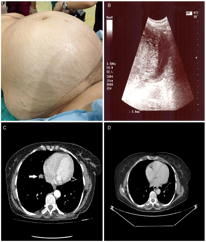
Fig. 1 (A) Giant invasive mole (28×25×15 cm) presenting with abdominopelvic mass. (B) Ultrasound images show a huge molar mass and marked thinning of the uterine wall. (C) The preoperative pulmonary computed tomography scan revealed metastatic nodules (white arrow), especially in basal segments of the right lung. (D) All of the nodules showed significant regression at 12th month after chemotherapy.
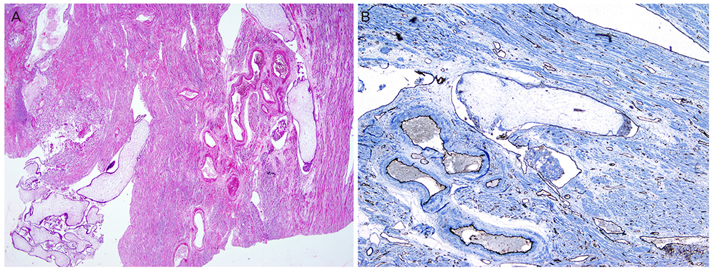
Fig. 2 (A) Distended hydropic villi and trophoblastic cells which invade into the adjacent myometrium (hematoxylin-eosin, ×40). (B) Lymphatic vessels with immunostaining of CD31 (immunoperoxidase, ×100).
Reference
-
1. Ozalp SS, Telli E, Oge T, Tulunay G, Boran N, Turan T, et al. Multicenter analysis of gestational trophoblastic neoplasia in Turkey. Asian Pac J Cancer Prev. 2014; 15:3625–3628.2. Lurain JR. Gestational trophoblastic disease I: epidemiology, pathology, clinical presentation and diagnosis of gestational trophoblastic disease, and management of hydatidiform mole. Am J Obstet Gynecol. 2010; 203:531–539.3. Hol K, Junnare K, Shekhawat GS, Damle H. A case of invasive mole with uterine rupture presenting as a hemoperitoneum. Glob J Res Anal. 2013; 2:195–196.4. Hurteau JA. Gestational trophoblastic disease: management of hydatidiform mole. Clin Obstet Gynecol. 2003; 46:557–569.5. Allen SD, Lim AK, Seckl MJ, Blunt DM, Mitchell AW. Radiology of gestational trophoblastic neoplasia. Clin Radiol. 2006; 61:301–313.6. Gibbs RS, Danforth DN, editors. Danforth's obstetrics and gynecology. 10th ed. Philadelphia (PA): Lippincott Williams & Wilkins;2008.7. Taskin S, Cengiz B, Ortac F. Invasive mole in a postmenopausal woman. Int J Gynaecol Obstet. 2006; 93:156–157.8. Bruner DI, Pritchard AM, Clarke J. Uterine rupture due to invasive metastatic gestational trophoblastic neoplasm. West J Emerg Med. 2013; 14:444–447.9. Kumar S, Vimala N, Mittal S. Invasive mole presenting as acute haemoperitoneum. JK Sci J Med Educ Res. 2004; 6:159–160.10. Seckl MJ, Sebire NJ, Fisher RA, Golfier F, Massuger L, Sessa C, et al. Gestational trophoblastic disease: ESMO Clinical Practice Guidelines for diagnosis, treatment and follow-up. Ann Oncol. 2013; 24:Suppl 6. vi39–vi50.11. Lok CA, Zurcher AF, van der Velden J. A case of a hydatidiform mole in a 56-year-old woman. Int J Gynecol Cancer. 2005; 15:163–166.12. von Welser SF, Grube M, Ortmann O. Invasive mole in a perimenopausal woman: a case report and systematic review. Arch Gynecol Obstet. 2015; 292:1193–1199.13. de la Fouchardiere A, Cassignol A, Benkiran L, Rudigoz RC, Gougeon A, Devouassoux-Shisheboran M. Invasive hydatiform mole in a postmenopausal woman. Ann Pathol. 2003; 23:443–446.14. Tsukamoto N, Iwasaka T, Kashimura Y, Uchino H, Kashimura M, Matsuyama T. Gestational trophoblastic disease in women aged 50 or more. Gynecol Oncol. 1985; 20:53–61.15. Bandy LC, Clarke-Pearson DL, Hammond CB. Malignant potential of gestational trophoblastic disease at the extreme ages of reproductive life. Obstet Gynecol. 1984; 64:395–399.
- Full Text Links
-
- Actions
-
Cited
- CITED
-
- Close
- Share
-
- Similar articles
-
- A Case of Invasive Mole with Uterine Rupture Presenting as a Hemoperitoneum
- A Case of Invasive Mole Initially Presenting with Symptoms of Brain Metastasis
- Metastatic gestational trophoblastic neoplasm presenting as spontaneous renal and cerebral hemorrhage with low titer of HCG: A case report of an unusual case
- Metastatic Invasive Mole in the Lung Arising from a Cornual Pregnancy
- Invasine Ductal Carcinoma with Osteoclast-Like Giant Cell in a Young Woman
