Analysis of the Effects of Different Iodine Concentrations on the Characterization of Small Renal Lesions Detected by Multidetector Computed Tomography Scan: A Pilot Study
- Affiliations
-
- 1Department of Radiology, Seoul National University College of Medicine, Seoul, Korea. kimshrad@snu.ac.kr
- 2Kidney Research Institute, Seoul National University College of Medicine, Seoul, Korea.
- 3Institute of Radiation Medicine, Seoul National University Medical Research Center, Seoul, Korea.
- 4Department of Radiology, Seoul National University Bundang Hospital, Seongnam, Korea.
- 5Clinical Research Institute, Seoul National University Hospital, Seoul, Korea.
- 6Department of Radiology, Seoul National University Hospital, Seoul, Korea.
- 7Department of Radiology, SMG-SNU Boramae Medical Center, Seoul, Korea.
- KMID: 2377037
- DOI: http://doi.org/10.3348/jksr.2017.76.5.337
Abstract
- PURPOSE
Our objective was to compare the effects of different iodine concentrations on characterizing small renal lesions.
MATERIALS AND METHODS
Thirty-eight patients were enrolled in this study. All patients underwent an initial CT scan using 370 mgI/mL iodinated contrast media. Patients were then randomized into two groups for a follow-up CT. Group A (n = 19) received 250 mgI/mL iodinated contrast media, and group B (n = 19) received 300 mgI/mL contrast media. The mean Hounsfield units (HU values) of small renal lesions with a maximum size of less than 2 cm were calculated. Signal to noise ratios (SNR values) were likewise evaluated. Three uroradiologists assessed the lesion's conspicuity and the diagnostic influence of the artifact's proximity to the adjacent renal parenchyma.
RESULTS
In group A, there were significant differences between the HU values of renal lesions and those of the adjacent renal parenchyma between the initial and follow-up CT. Conversely, in group B, there was no significant difference. Moreover, SNR values showed no statistically significant difference between both groups. Regarding lesion conspicuity, only one reader identified a significant difference (p = 0.032) in group A; whereas in group B, there was no statistical difference. The artifact's proximity to the adjacent renal parenchyma did not appear to have any diagnostic influence on differentiating the two (p < 0.05).
CONCLUSION
In evaluating small renal lesions, 300 mgI/mL instead of 370 mgI/mL contrast media can be used; however, it is important to note that the use of 250 mgI/mL contrast media may reveal different results from that of 370 mgI/mL contrast media.
MeSH Terms
Figure
-
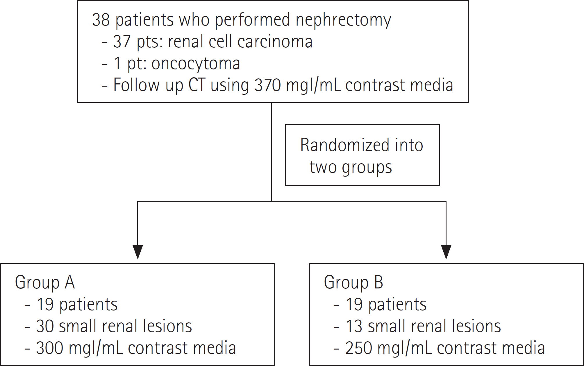
Fig. 1. Flow chart demonstrating the included patients. 38 patients underwent CT examination after injection of 370 mgI/mL contrast media. Subsequently, for the followup CT, the patients were randomized into two groups. Group A (n = 19) underwent their followup CT after injection of 250 mgI/mL contrast media, and group B (n = 19) underwent CT examination after injection of 300 mgI/mL contrast media.
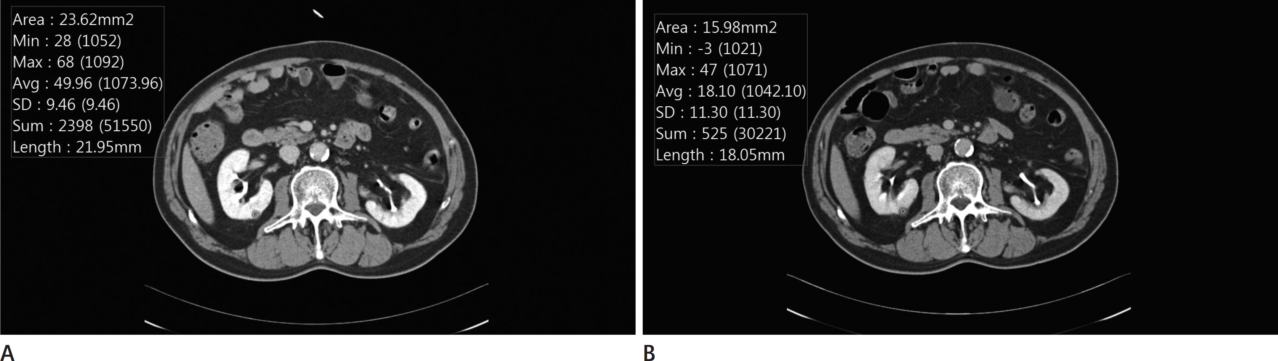
Fig. 2. Small renal lesion of a 54-year-old male patient; imaged using 370 mgI/mL (A) and 250 mgI/mL contrast medium (B). A. The HU of the small renal lesion, imaged with 370 mgI/mL of contrast medium, was 50. B. The HU of the same lesion, imaged with 250 mgI/mL of contrast medium, was 18.1. There was a significant difference in the HUs of the small renal lesions in group A. HU = Hounsfield unit
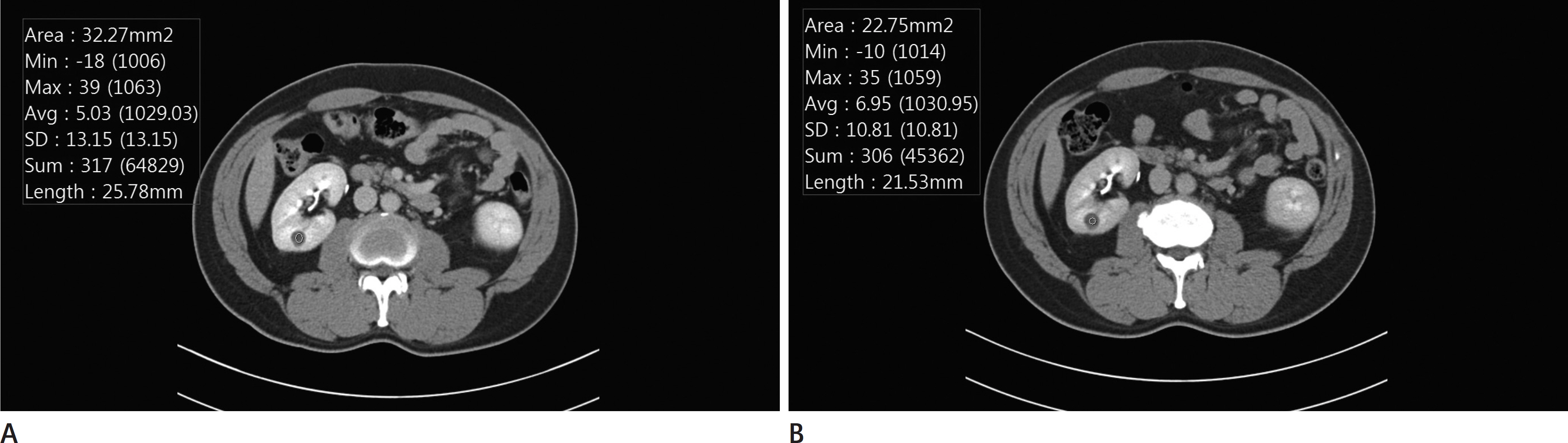
Fig. 3. A small renal lesion of a 41-year-old male patient; imaged using 370 mgI/mL (A) and 300 mgI/mL of contrast medium (B). A. The HU of the small renal lesion, imaged with 370 mgI/mL of contrast medium, was 5.0. B. The HU of the same lesion, imaged with 300 mgI/mL of contrast medium, was 7.0. There was no significant difference in the HUs of the small renal lesions in group B. HU = Hounsfield unit
Reference
-
1. Rengo M, Caruso D, De Cecco CN, Lucchesi P, Bellini D, Ma-ceroni MM, et al. High concentration (400 mgI/mL) versus low concentration (320 mgI/mL) iodinated contrast media in multi detector computed tomography of the liver: a randomized, single centre, noninferiority study. Eur J Radiol. 2012; 81:3096–3101.2. Rengo M, Bellini D, De Cecco CN, Osimani M, Vecchietti F, Caruso D, et al. The optimal contrast media policy in CT of the liver. Part II: clinical protocols. Acta Radiol. 2011; 52:473–480.
Article3. Laghi A. Multidetector CT (64 slices) of the liver: examination techniques. Eur Radiol. 2007; 17:675–683.
Article4. Marchianò A, Spreafico C, Lanocita R, Frigerio L, Di Tolla G, Patelli G, et al. Does iodine concentration affect the diagnostic efficacy of biphasic spiral CT in patients with hepatocellular carcinoma? Abdom Imaging. 2005; 30:274–280.
Article5. Kondo H, Kanematsu M, Goshima S, Tomita Y, Kim MJ, Moriyama N, et al. Body size indexes for optimizing iodine dose for aortic and hepatic enhancement at multidetector CT: comparison of total body weight, lean body weight, and blood volume. Radiology. 2010; 254:163–169.
Article6. Kondo H, Kanematsu M, Goshima S, Watanabe H, Onozuka M, Moriyama N, et al. Aortic and hepatic enhancement at multidetector CT: evaluation of optimal iodine dose deter-mined by lean body weight. Eur J Radiol. 2011; 80:e273–e277.
Article7. Yanaga Y, Awai K, Nakaura T, Namimoto T, Oda S, Funama Y, et al. Optimal contrast dose for depiction of hypervas-cular hepatocellular carcinoma at dynamic CT using 64-MDCT. AJR Am J Roentgenol. 2008; 190:1003–1009.
Article8. Fleischmann D. Multiple detector-row CT angiography of the renal and mesenteric vessels. Eur J Radiol. 2003; 45(Suppl 1):S79–S87.
Article9. Johnson PT, Fishman EK. IV contrast selection for MDCT: current thoughts and practice. AJR Am J Roentgenol. 2006; 186:406–415.
Article10. Loewe C, Becker CR, Berletti R, Cametti CA, Caudron J, Cou-dyzer W, et al. 64-Slice CT angiography of the abdominal aorta and abdominal arteries: comparison of the diagnostic efficacy of iobitridol 350 mgI/mL versus iomeprol 400 mgI/mL in a prospective, randomised, double-blind multi-centre trial. Eur Radiol. 2010; 20:572–583.11. Katzberg RW, Barrett BJ. Risk of iodinated contrast material–induced nephropathy with intravenous administration. Radiology. 2007; 243:622–628.12. Thomsen HS, Morcos SK, Barrett BJ. Contrast-induced ne-phropathy: the wheel has turned 360 degrees. Acta Radiol. 2008; 49:646–657.
Article13. Nyman U, Almén T, Aspelin P, Hellström M, Kristiansson M, Sterner G. Contrast-medium-Induced nephropathy corre-lated to the ratio between dose in gram iodine and esti-mated GFR in mL/min. Acta Radiol. 2005; 46:830–842.
Article14. Lim A, O'Neil B, Heilbrun ME, Dechet C, Lowrance WT. The contemporary role of renal mass biopsy in the management of small renal tumors. Front Oncol. 2012; 2:106.
Article15. Lee CT, Katz J, Shi W, Thaler HT, Reuter VE, Russo P. Surgical management of renal tumors 4 cm. Or less in a contemporary cohort. J Urol. 2000; 163:730–736.
Article16. Nguyen MM, Gill IS, Ellison LM. The evolving presentation of renal carcinoma in the United States: trends from the surveillance, epidemiology, and end results program. J Urol. 2006; 176(6 Pt 1):2397–2400. discussion. 2400.
Article17. Alasker A, Williams SK, Ghavamian R. Small renal mass: to treat or not to treat. Curr Urol Rep. 2013; 14:13–18.
Article18. Sandstede JJ, Kaupert C, Roth A, Jenett M, Harz C, Hahn D. Comparison of different iodine concentrations for multidetector row computed tomography angiography of segmen-tal renal arteries. Eur Radiol. 2005; 15:1211–1214.
Article19. Sandstede JJ, Werner A, Kaupert C, Roth A, Jenett M, Harz C, et al. A prospective study comparing different iodine concentrations for triphasic multidetector row CT of the upper abdomen. Eur J Radiol. 2006; 60:95–99.
Article20. Kaufman L, Kramer DM, Crooks LE, Ortendahl DA. Measur-ing signal-to-noise ratios in MR imaging. Radiology. 1989; 173:265–267.
Article21. Firbank MJ, Coulthard A, Harrison RM, Williams ED. A comparison of two methods for measuring the signal to noise ratio on MR images. Phys Med Biol. 1999; 44:N261–N264.
Article22. Magnotta VA, Friedman L. FIRST BIRN. Measurement of signal-to-noise and contrast-to-noise in the fBIRN multicenter imaging study. J Digit Imaging. 2006; 19:140–147.
Article23. Setty BN, Sahani DV, Ouellette-Piazzo K, Hahn PF, Shepard JA. Comparison of enhancement, image quality, cost, and adverse reactions using 2 different contrast medium concentrations for routine chest CT on 16-slice MDCT. J Comput Assist Tomogr. 2006; 30:818–822.
Article24. Holalkere NS, Matthes K, Kalva SP, Brugge WR, Sahani DV. 64-slice multidetector row CT angiography of the abdomen: comparison of low versus high concentration iodinated contrast media in a porcine model. Br J Radiol. 2011; 84:221–228.
Article25. Kundel HL, Polansky M. Measurement of observer agree-ment. Radiology. 2003; 228:303–308.
Article26. Bae KT. Optimization of contrast enhancement in thoracic MDCT. Radiol Clin North Am. 2010; 48:9–29.
Article27. Schoellnast H, Brader P, Oberdabernig B, Pisail B, Deut-schmann HA, Fritz GA, et al. High-concentration contrast media in multiphasic abdominal multidetector-row computed tomography: effect of increased iodine flow rate on parenchymal and vascular enhancement. J Comput Assist Tomogr. 2005; 29:582–587.28. Berland LL, Lee JY. Comparison of contrast media injection rates and volumes for hepatic dynamic incremented computed tomography. Invest Radiol. 1988; 23:918–922.
Article29. Chambers TP, Baron RL, Lush RM. Hepatic CT enhancement. Part I. Alterations in the volume of contrast material within the same patients. Radiology. 1994; 193:513–517.
Article30. Dean PB, Violante MR, Mahoney JA. Hepatic CT contrast enhancement: effect of dose, duration of infusion, and time elapsed following infusion. Invest Radiol. 1980; 15:158–161.
Article31. Heiken JP, Brink JA, McClennan BL, Sagel SS, Crowe TM, Gaines MV. Dynamic incremental CT: effect of volume and concentration of contrast material and patient weight on hepatic enhancement. Radiology. 1995; 195:353–357.
Article32. Heiken JP, Brink JA, McClennan BL, Sagel SS, Forman HP, DiCroce J. Dynamic contrast-enhanced CT of the liver: comparison of contrast medium injection rates and uniphasic and biphasic injection protocols. Radiology. 1993; 187:327–331.
Article33. Ramgren B, Björkman-Burtscher IM, Holtås S, Siemund R. CT angiography of intracranial arterial vessels: impact of tube voltage and contrast media concentration on image quality. Acta Radiol. 2012; 53:929–934.
Article34. Bagrodia A, Darwish OM, Rapoport Y, Margulis V. Risk pre-diction in the management of small renal masses. Curr Opin Urol. 2012; 22:347–352.
Article35. Gill IS, Aron M, Gervais DA, Jewett MA. Clinical practice. Small renal mass. N Engl J Med. 2010; 362:624–634.36. Volpe A, Panzarella T, Rendon RA, Haider MA, Kondylis FI, Jewett MA. The natural history of incidentally detected small renal masses. Cancer. 2004; 100:738–745.
Article
- Full Text Links
-
- Actions
-
Cited
- CITED
-
- Close
- Share
-
- Similar articles
-
- A Comparison of the Use of Contrast Media with Different Iodine Concentrations for Multidetector CT of the Kidney
- Characterization of Small Renal Masses Less than 4 cm with Quadriphasic Multidetector Helical Computed Tomography: Differentiation of Benign and Malignant Lesions
- Comparison of enhancement and image quality: different iodine concentrations for liver on 128-slice multidetector computed tomography in the same chronic liver disease patients
- Periureteral Varices with Accompanying Pyelitis Diagnosed by 3-Dimensional Reformatted Technique of the Multidetector Row CT: A Case Report
- Transient Orbitofacial Angioedema due to Intravenous Iodinated Contrast Media During Computed Tomography: CT Findings
