J Korean Assoc Oral Maxillofac Surg.
2017 Apr;43(2):77-82. 10.5125/jkaoms.2017.43.2.77.
Mechanical evaluation of the use of conventional and locking miniplate/screw systems used in sagittal split ramus osteotomy
- Affiliations
-
- 1Division of Oral and Maxillofacial Surgery, State University of Campinas, Campinas, Brazil.
- 2Division of Oral and Maxillofacial Surgery & CEMYQ, Universidad de La Frontera, Temuco, Chile. sergio.olate@ufrontera.cl
- 3Center for Biomedical Research, Universidad Autónoma de Chile, Temuco, Chile.
- KMID: 2377009
- DOI: http://doi.org/10.5125/jkaoms.2017.43.2.77
Abstract
OBJECTIVES
The aim of this study was to compare the mechanical resistance of four different osteosyntheses modeled in two different sagittal split ramus osteotomy (SSRO) designs and to determine the linear loading in a universal testing machine.
MATERIALS AND METHODS
An in vitro experiment was conducted with 40 polyurethane hemimandibles. The samples were divided into two groups based on osteotomy design; Group I, right angles between osteotomies and Group II, no right angles between osteotomies. In each group, the hemimandibles were distributed into four subgroups according to the osteosynthesis method, using one 4-hole 2.0 mm conventional or locking plate, with or without one bicortical screw with a length of 12.0 mm (hybrid technique). Each subgroup contained five samples and was subjected to a linear loading test in a universal testing machine.
RESULTS
The peak load and peak displacement were compared for statistical significance using PASW Statistics 18.0 (IBM Co., USA). In general, there was no difference between the peak load and peak displacement related to osteotomy design. However, when the subgroups were compared, the osteotomy without right angles offered higher mechanical resistance when one conventional or locking 2.0 mm plate was used. One locking plate with one bicortical screw showed higher mechanical resistance (162.72±42.55 N), and these results were statistically significantly compared to one conventional plate with monocortical screws (P=0.016) and one locking plate with monocortical screws (P=0.012). The difference in peak displacement was not statistically significant based on osteotomy design or internal fixation system configuration.
CONCLUSION
The placement of one bicortical screw in the distal region promoted better stabilization of SSRO. The osteotomy design did not influence the mechanical behavior of SSRO when the hybrid technique was applied.
MeSH Terms
Figure
-
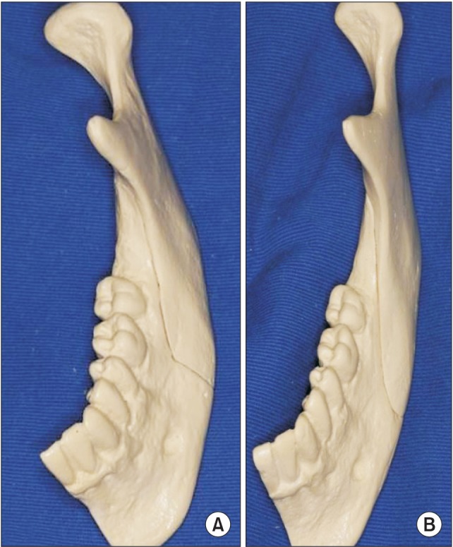
Fig. 1 Polyurethane hemimandibles according to sagittal split ramus osteotomy design. A. Group I, right angles between osteotomies. B. Group II, no angles between osteotomies.
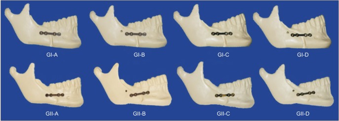
Fig. 2 GI, linear osteotomy; GII, angular osteotomy. Subgroups: A, one 4-hole 2.0 mm plate; B, one 4-hole 2.0 mm plate and one bicortical screw; C, one 4-hole 2.0 mm locking plate; D, one 4-hole 2.0 mm locking plate and one bicortical screw.
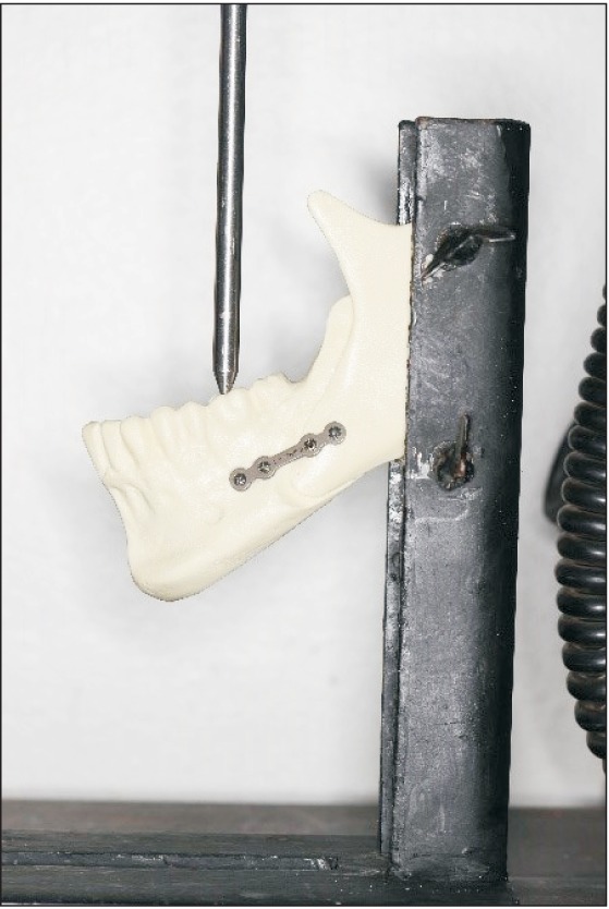
Fig. 3 Hemimandible in a metallic iron alloy support subjected to linear loading in a servohydraulic material testing unit (Instron 4411; Instron Corp., USA).
Reference
-
1. Böckmann R, Meyns J, Dik E, Kessler P. The modifications of the sagittal ramus split osteotomy: a literature review. Plast Reconstr Surg Glob Open. 2015; 2:e271. PMID: 25587505.2. Dal Pont G. Retro-molar osteotomy for correction of prognathism. Minerva Chir. 1959; 14:1138–1141. PMID: 13813774.3. Nishioka GJ, Aragon SB. Modified sagittal split technique for patients with a high lingula. J Oral Maxillofac Surg. 1989; 47:426–427. PMID: 2926556.
Article4. Ueki K, Okabe K, Miyazaki M, Mukozawa A, Marukawa K, Nakagawa K, et al. Position of mandibular canal and ramus morphology before and after sagittal split ramus osteotomy. J Oral Maxillofac Surg. 2010; 68:1795–1801. PMID: 20044190.
Article5. Chuong CJ, Borotikar B, Schwartz-Dabney C, Sinn DP. Mechanical characteristics of the mandible after bilateral sagittal split ramus osteotomy: comparing 2 different fixation techniques. J Oral Maxillofac Surg. 2005; 63:68–76. PMID: 15635560.
Article6. Wolford LM, Bennett MA, Rafferty CG. Modification of the mandibular ramus sagittal split osteotomy. Oral Surg Oral Med Oral Pathol. 1987; 64:146–155. PMID: 3476891.
Article7. Turvey TA, Bell RB, Tejera TJ, Proffit WR. The use of self-reinforced biodegradable bone plates and screws in orthognathic surgery. J Oral Maxillofac Surg. 2002; 60:59–65. PMID: 11757010.
Article8. Oguz Y, Uckan S, Ozden AU, Uckan E, Eser A. Stability of locking and conventional 2.0-mm miniplate/screw systems after sagittal split ramus osteotomy: finite element analysis. Oral Surg Oral Med Oral Pathol Oral Radiol Endod. 2009; 108:174–177. PMID: 19615655.
Article9. Chiodo TA, Ziccardi VB, Janal M, Sabitini C. Failure strength of 2.0 locking versus 2.0 conventional Synthes mandibular plates: a laboratory model. J Oral Maxillofac Surg. 2006; 64:1475–1479. PMID: 16982304.
Article10. Pozzer L, Olate S, Cavalieri-Pereira L, de Moraes M, Albergaría-Barbosa JR. Influence of the design in sagittal split ramus osteotomy on the mechanical behavior. Int J Clin Exp Med. 2014; 7:1284–1288. PMID: 24995084.11. Trauner R, Obwegeser H. The surgical correction of mandibular prognathism and retrognathia with consideration of genioplasty. I. Surgical procedures to correct mandibular prognathism and reshaping of the chin. Oral Surg Oral Med Oral Pathol. 1957; 10:677–689. PMID: 13441284.12. Haug RH. Effect of screw number on reconstruction plating. Oral Surg Oral Med Oral Pathol. 1993; 75:664–668. PMID: 8515976.
Article13. Gutwald R, Alpert B, Schmelzeisen R. Principle and stability of locking plates. Keio J Med. 2003; 52:21–24. PMID: 12713018.
Article14. Ribeiro-Junior PD, Magro-Filho O, Shastri KA, Papageorge MB. Which kind of miniplate to use in mandibular sagittal split osteotomy? An in vitro study. Int J Oral Maxillofac Surg. 2012; 41:1369–1373. PMID: 22658672.
Article15. Ribeiro-Junior PD, Magro-Filho O, Shastri KA, Papageorge MB. In vitro biomechanical evaluation of the use of conventional and locking miniplate/screw systems for sagittal split ramus osteotomy. J Oral Maxillofac Surg. 2010; 68:724–730. PMID: 19962812.
Article16. Schwartz HC, Relle RJ. Bicortical-monocortical fixation of the sagittal mandibular osteotomy. J Oral Maxillofac Surg. 1996; 54:234–235. PMID: 8604080.
Article17. Sato FR, Asprino L, Consani S, Noritomi PY, de Moraes M. A comparative evaluation of the hybrid technique for fixation of the sagittal split ramus osteotomy in mandibular advancement by mechanical, photoelastic, and finite element analysis. Oral Surg Oral Med Oral Pathol Oral Radiol. 2012; 114(5 Suppl):S60–S68. PMID: 23083958.
Article18. Andrade VC, Olate S, Pozzer L, Cavalieri-Pereira L, de Moraes M, de Albergaria-Barbosa JR. Photoelastic evaluation of two different sagittal split ramus osteotomies in advancement surgery. Int J Clin Exp Med. 2014; 7:1940–1944. PMID: 25232374.
- Full Text Links
-
- Actions
-
Cited
- CITED
-
- Close
- Share
-
- Similar articles
-
- Treatment of osteomyelitis in the rear area of the lingula of the mandible using sagittal split ramus osteotomy: a case report
- A case report of hemifacial microsomia
- Facial palsy after sagittal split ramus osteotomies: Case report
- Enucleation of large keratocystic odontogenic tumor at mandible via unilateral sagittal split osteotomy: a report of three cases
- Comparative study of stability and relapse according to fixation method after bilateral sagittal split ramus osteotomies in mandibular prognathic patients
