Prog Med Phys.
2016 Sep;27(3):125-130. 10.14316/pmp.2016.27.3.125.
Optimization of Energy Modulation Filter for Dual Energy CBCT Using Geant4 Monte-Carlo Simulation
- Affiliations
-
- 1Department of Medical Science, Ewha Womans University, Korea.
- 2Department of Radiation Oncology, School of Medicine, Yonsei University, Seoul, Korea.
- 3Department of Radiation Oncology, School of Medicine, Dankook University, Cheonan, Korea.
- 4Department of Biomedical Engineering, School of Medicine, Ewha Womans University, Seoul, Korea. renalee@ewha.ac.kr
- KMID: 2376542
- DOI: http://doi.org/10.14316/pmp.2016.27.3.125
Abstract
- Dual energy computed tomography (DECT) is used to classify two materials and quantify the mass density of each material in the human body. An energy modulation filter based DECT could acquire two images, which are generated by the low- and high-energy photon spectra, in one scan, with one tube and detector. In the case of DECT using the energy modulation filter, the filter should perform the optimization process for the type of materials and thicknesses for generating two photon spectra. In this study, Geant4 Monte-Carlo simulation toolkit was used to execute the optimization process for determining the property of the energy modulation filter. In the process, various materials used for the energy modulation filter are copper (Cu, 8.96 g/cm³), niobium (Nb, 8.57 g/cm³), stannum (Sn, 7.31 g/cm³), gold (Au, 19.32 g/cm³), and lead (Pb, 11.34 g/cm³). The thickness of the modulation filter varied from 0.1 mm to 1.0 mm. To evaluate the overlap region of the low- and high-energy spectrum, Geant4 Monte-Carlo simulation is used. The variation of the photon flux and the mean energy of photon spectrum that passes through the energy modulation filter are evaluated. In the primary photon spectrum of 80 kVp, the optimal modulation filter is a 0.1 mm lead filter that can acquire the same mean energy of 140 kVp photon spectrum. The lead filter of 0.1 mm based dual energy CBCT is required to increase the tube current 4.37 times than the original tube current owing to the 77.1% attenuation in the filter.
Figure
-
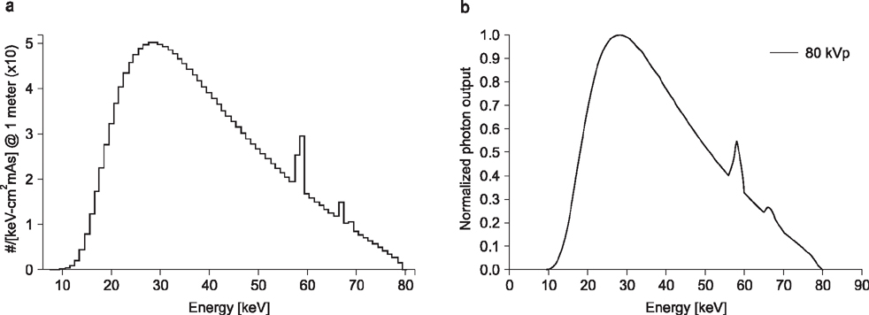
Fig. 1 80kVp Photon spectrum. (a) The incident photon spectrum was created by the SpekCalc program and applied to the simulation, (b) the photon spectrum was measured by Geant4.
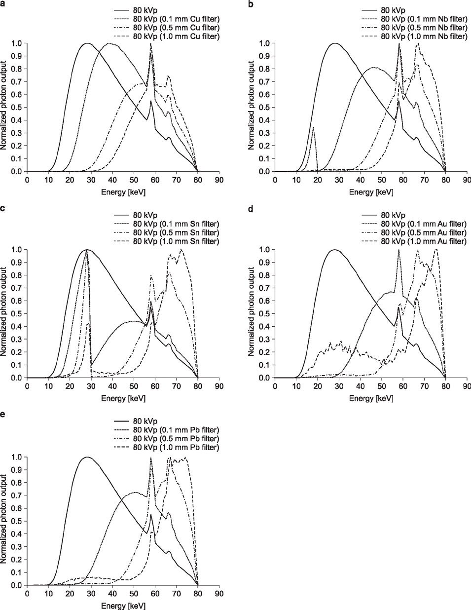
Fig. 2 80 kVp spectrum change by the thickness of energy modulation filter from 0.1 to 1 mm for each material. (a) Copper, (b) Niobium, (c) Stannum, (d) Gold, (e) Lead.
Cited by 1 articles
-
Estimation of Noise Level and Edge Preservation for Computed Tomography Images: Comparisons in Iterative Reconstruction
Sihwan Kim, Chulkyun Ahn, Woo Kyoung Jeong, Jong Hyo Kim, Minsoo Chun
Prog Med Phys. 2021;32(4):92-98. doi: 10.14316/pmp.2021.32.4.92.
Reference
-
1. Primak AN, Giraldo JCR, Liu X, et al. Improved dual energy material discrimination for dual source CT by means of additional spectral filtration. Med Phys. 2009; 36(4):1359–1369.2. Landry G, et al. Extracting atomic numbers and electron densities from a dual source dual energy CT scanner: experiments and a simulation model. Radiother Oncol. 2011; 100(3):375–379.
Article3. Johnson TR. Dual energy CT: general principles. AJR Am J Roentgenol. 2012; 199:S3–S8.4. Petersilka M, Bruder H, Krauss B, Stierstorfer K, Flohr TG. Technical principles of dual source CT. Eur J Radiol. 2008; 68(3):362–368.
Article5. Altman A, Carmi R. A double-layer detector, dual-energy CT —principles, advantages and applications. Med Phys. 2009; 36(6):2750–2750.6. Saito M. Spectral optimization for measuring electron density by the dual energy computed tomography coupled with balanced filter method. Med Phys. 2009; 36(8):3631–3642.7. Nasseri MM. Determination of tungsten target parameters for transmission X-ray tube: A simulation study using Geant4. Nuc Eng Technol. 2016; 48:795–798.8. Agostinelli S, Allison J, Amako KA, et al. GEANT4—a simulation toolkit. Nuclear Instruments and Methods in Physics Research A. 2003; 506(3):250–303.9. Guthoff M, et al. Geant4 simulation of a filtered X-ray source for radiation damage studies. Nuclear Instruments and Methods in Physics Research Section A: Accelerators, Spectrometers, Detectors and Associated Equipmen. 2012; 675:118–122.
Article10. Poludniowski G, et al. SpekCalc: a program to calculate photon spectra from tungsten anode X-ray tubes. Phys Med Biol. 2009; 54(19):N433.
Article11. Bazalova M, et al. Tissue segmentation in Monte Carlo treatment planning: a simulation study using dual-energy CT images. Radiother Oncol. 2008; 86(1):93–98.
Article12. Danad I, Fayad ZA, Willemink MJ, Min JK. New applications of cardiac computed tomography: dual-energy, spectral, and molecular CT imaging. JACC Cardiovasc Imaging. 2015; 8(6):710–723.13. Johnson T. Dual Energy CT in Clinical Practice. Springer Science & Business Media;2011. p. 3–8.
- Full Text Links
-
- Actions
-
Cited
- CITED
-
- Close
- Share
-
- Similar articles
-
- Analysis of Beam Hardening of Modulation Layers for Dual Energy Cone-beam CT
- Study on the 6 MV Photon Beam Characteristics and Analysis Method from Medical Linear Accelerators Using Geant4 Medical Linac2 Example
- A Monte Carlo Simulation Study of a Therapeutic Proton Beam Delivery System Using the Geant4 Code
- Evaluation of Radiation Dose for Dual Energy CBCT Using Multi-Grid Device
- Development of DICOM Convert Program for the Geant4 Monte Carlo Simulation of the Radiotherapy
