Laminar Cortical Hypointensities in Susceptibility-Weighted Imaging in a Case of Progressive Multifocal Leukoencephalopathy
- Affiliations
-
- 1Second Department of Neurology, Attikon Hospital, School of Medicine, National and Kapodistrian University of Athens, Athens, Greece. tsivgoulisgiorg@yahoo.gr
- 2Iatropolis Magnetic Resonance Diagnostic Centre, Athens, Greece.
- 3Stroke Unit, Metropolitan Hospital, Piraeus, Greece.
- 4Department of Neurology, The University of Tennessee Health Science Center, Memphis, TN, USA.
- 5International Clinical Research Center, Department of Neurology, St. Anne's University Hospital in Brno, Brno, Czech Republic.
- KMID: 2376024
- DOI: http://doi.org/10.3988/jcn.2017.13.2.201
Abstract
- No abstract available.
MeSH Terms
Figure
-
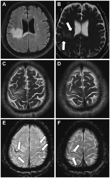
Fig. 1 Three Tesla Brain MRI findings. A: Brain FLAIR sequence showing hyperintense lesions in the U-fibers, deep white matter of the right hemisphere, and the splenium of the corpus callosum. B: Brain apparent diffusion coefficient sequence showing restricted diffusivity at the lesion borders (arrows), which is a characteristic neuroimaging finding of PML. C and D: T2-weighted MRI sequence showing sparing of the cortex at the involved right precentral and postcentral gyri. E and F: SWI images revealing laminar hypointensities of the cortex (arrows) at the involved sites and also over normal-appearing white matter. FLAIR: fluid attenuated inversion recovery, PML: progressive multifocal leukoencephalopathy, SWI: susceptibility-weighted imaging.
Reference
-
1. Wattjes MP, Richert ND, Killestein J, de Vos M, Sanchez E, Snaebjornsson P, et al. The chameleon of neuroinflammation: magnetic resonance imaging characteristics of natalizumab-associated progressive multifocal leukoencephalopathy. Mult Scler. 2013; 19:1826–1840.
Article2. Berger JR, Aksamit AJ, Clifford DB, Davis L, Koralnik IJ, Sejvar JJ, et al. PML diagnostic criteria: consensus statement from the AAN Neuroinfectious Disease Section. Neurology. 2013; 80:1430–1438.3. Dong-Si T, Richman S, Wattjes MP, Wenten M, Gheuens S, Philip J, et al. Outcome and survival of asymptomatic PML in natalizumab-treated MS patients. Ann Clin Transl Neurol. 2014; 1:755–764.
Article4. Labauge P, Carra-Dalliere C, Ayrignac X, Menjot de Champfleur N. Low signals on T2* and SWI sequences in patients with MS with progressive multifocal leukoencephalopathy. AJNR Am J Neuroradiol. 2016; 37:E11.5. Carra-Dalliere C, Menjot de Champfleur N, Deverdun J, Ayrignac X, Nerrant E, Makinson A, et al. Use of quantitative susceptibility mapping (QSM) in progressive multifocal leukoencephalopathy. J Neuroradiol. 2016; 43:6–10.
Article6. Umino M, Maeda M, Ii Y, Tomimoto H, Sakuma H. Low-signal-intensity rim on susceptibility-weighted imaging is not a specific finding to progressive multifocal leukoencephalopathy. J Neurol Sci. 2016; 362:155–159.
Article
- Full Text Links
-
- Actions
-
Cited
- CITED
-
- Close
- Share
-
- Similar articles
-
- Hypointense Rim on Susceptibility-Weighted Imaging in a Patient with Progressive Multifocal Leukoencephalopathy
- Progressive Multifocal Leukoencephalopathy in AIDS: Proton MR Spectroscopy Patterns of Asynchronous Lesions Confirmed by Serial Diffusion-Weighted Imaging and Apparent Diffusion Coefficient Mapping
- Recurrent Complex Partial Seizures in a Patient with Progressive Multifocal Leukoencephalopathy
- Progressive Multifocal Leukoencephalopathy Presenting as Viral Encephalitis in a Healthy Adult
- Cortical Laminar Necrosis in an Infant with Severe Traumatic Brain Injury
