Medical Thoracoscopy in Pleural Disease: Experience from a One-Center Study
- Affiliations
-
- 1Division of Pulmonary and Critical Care Medicine, Department of Internal Medicine, Ewha Womans University School of Medicine, Seoul, Korea.
- 2Division of Pulmonary and Critical Care Medicine, Department of Internal Medicine, Seoul National University College of Medicine, Seoul, Korea. mdyspark@gmail.com
- KMID: 2375989
- DOI: http://doi.org/10.4046/trd.2017.80.2.194
Abstract
- BACKGROUND
Medical thoracoscopy (MT) is a minimally invasive, endoscopic procedure for exploration of the pleural cavity under conscious sedation and local anesthesia. MT has been performed at the Seoul National University Hospital since February 2014. This paper summarizes the findings and outcomes of MT cases at this hospital.
METHODS
Patients who had undergone MT were enrolled in the study. MT was performed by pulmonologists, using both rigid and semi-rigid thoracoscopes. During the procedure, patients were under conscious sedation with fentanyl and midazolam. Medical records were reviewed for clinical data.
RESULTS
From February 2014 to January 2016, 50 procedures (47 cases) were performed (diagnostic MT, 26 cases; therapeutic MT, 24 cases). The median age of patients was 66 years (59-73 years), and 38 patients (80.9%) were male. The median procedure duration from initial incision to insertion of the chest tube was 37 minutes. The median doses of fentanyl and midazolam were 50 µg and 5 mg, respectively. All procedures were performed without unexpected events. Of the 26 cases of pleural disease with an unknown cause, 19 were successfully diagnosed using MT. Additionally, diagnostic MT provided clinically useful information in the other six patients. Therapeutic MT was very effective for treatment of malignant pleural effusion or empyema. The median number of days with chest tube drainage was 6 (3 days for diagnostic MT and 8 days for therapeutic MT).
CONCLUSION
MT is a useful and necessary procedure for both diagnosis and treatment of pleural diseases.
Keyword
MeSH Terms
Figure
-
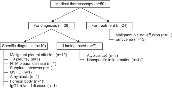
Figure 1 Flow diagram of patients undergoing medical thoracoscopy. *Guidewire. †Adenocarcinoma was diagnosed via video-assisted thoracoscopic surgery (VATS) in one patient; two patients were clinically diagnosed with malignant pleural effusion. ‡Mesothelioma was diagnosed via VATS in one patient, and pleural effusion was spontaneously resolved in another three patients. TB: tuberculosis; NTM: nontuberculous mycobacteria; GVHD: graft-versus-host disease.
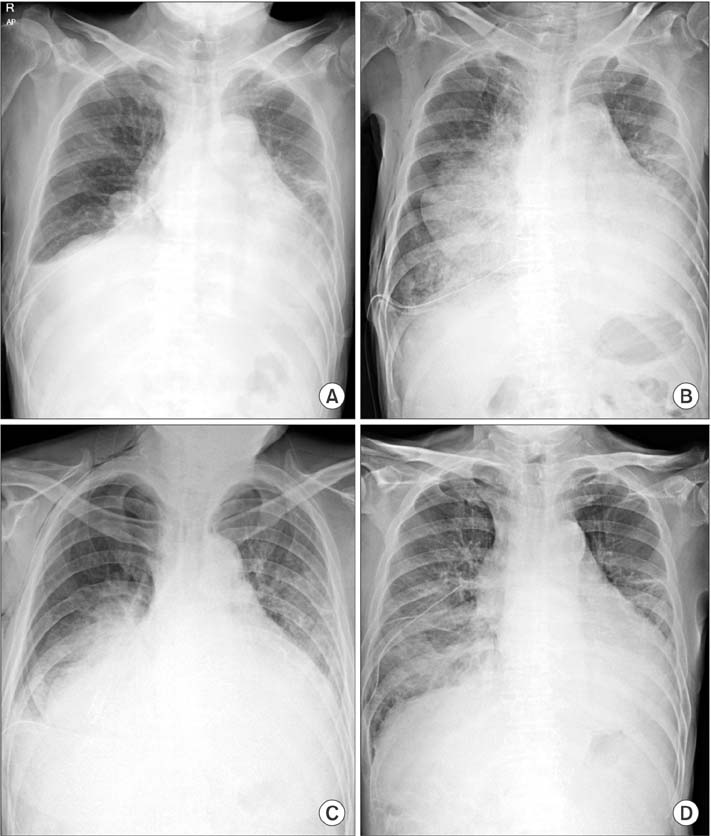
Figure 2 Re-expansion pulmonary edema. Chest radiography immediately (A), 4 hours (B), 6 hours (C), and 3 days (D) after medical thoracoscopy.
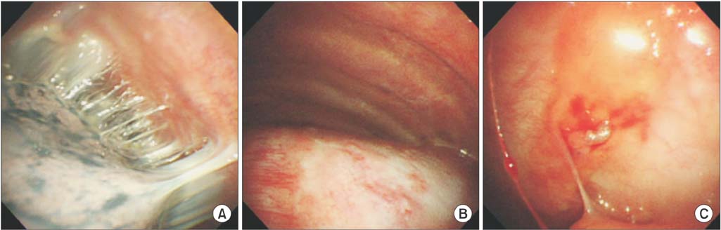
Figure 3 IgG4-related disease: findings during medical thoracoscopy. (A) Adhesions in the pleural space. (B) Thickened parietal pleura. (C) Inflamed parietal pleura with proliferating blood vessels.
Reference
-
1. Lee P, Mathur PN, Colt HG. Advances in thoracoscopy: 100 years since Jacobaeus. Respiration. 2010; 79:177–186.2. Michaud G, Berkowitz DM, Ernst A. Pleuroscopy for diagnosis and therapy for pleural effusions. Chest. 2010; 138:1242–1246.3. Rahman NM, Ali NJ, Brown G, Chapman SJ, Davies RJ, Downer NJ, et al. Local anaesthetic thoracoscopy: British Thoracic Society Pleural Disease Guideline 2010. Thorax. 2010; 65:Suppl 2. ii54–ii60.4. Agarwal R, Aggarwal AN, Gupta D. Diagnostic accuracy and safety of semirigid thoracoscopy in exudative pleural effusions: a meta-analysis. Chest. 2013; 144:1857–1867.5. Brutsche MH, Tassi GF, Gyorik S, Gokcimen M, Renard C, Marchetti GP, et al. Treatment of sonographically stratified multiloculated thoracic empyema by medical thoracoscopy. Chest. 2005; 128:3303–3309.6. Shojaee S, Lee HJ. Thoracoscopy: medical versus surgical-in the management of pleural diseases. J Thorac Dis. 2015; 7:Suppl 4. S339–S351.7. Migliore M, Giuliano R, Aziz T, Saad RA, Sgalambro F. Four-step local anesthesia and sedation for thoracoscopic diagnosis and management of pleural diseases. Chest. 2002; 121:2032–2035.8. Hersh CP, Feller-Kopman D, Wahidi M, Garland R, Herth F, Ernst A. Ultrasound guidance for medical thoracoscopy: a novel approach. Respiration. 2003; 70:299–301.9. Marchetti G, Valsecchi A, Indellicati D, Arondi S, Trigiani M, Pinelli V. Ultrasound-guided medical thoracoscopy in the absence of pleural effusion. Chest. 2015; 147:1008–1012.10. Havelock T, Teoh R, Laws D, Gleeson F. BTS Pleural Disease Guideline Group. Pleural procedures and thoracic ultrasound: British Thoracic Society Pleural Disease Guideline 2010. Thorax. 2010; 65:Suppl 2. ii61–ii76.11. Hallifax RJ, Corcoran JP, Ahmed A, Nagendran M, Rostom H, Hassan N, et al. Physician-based ultrasound-guided biopsy for diagnosing pleural disease. Chest. 2014; 146:1001–1006.12. Lee P, Hsu A, Lo C, Colt HG. Prospective evaluation of flex-rigid pleuroscopy for indeterminate pleural effusion: accuracy, safety and outcome. Respirology. 2007; 12:881–886.13. Dhooria S, Singh N, Aggarwal AN, Gupta D, Agarwal R. A randomized trial comparing the diagnostic yield of rigid and semirigid thoracoscopy in undiagnosed pleural effusions. Respir Care. 2014; 59:756–764.14. Willendrup F, Bodtger U, Colella S, Rasmussen D, Clementsen PF. Diagnostic accuracy and safety of semirigid thoracoscopy in exudative pleural effusions in Denmark. J Bronchology Interv Pulmonol. 2014; 21:215–219.15. Yang JK, Lee JH, Kwon MH, Jeong JH, Lee GE, Cho HM, et al. Diagnostic accuracy and safety of medical thoracoscopy. Tuberc Respir Dis. 2007; 63:261–267.16. Ishida A, Furuya N, Nishisaka T, Mineshita M, Miyazawa T. IgG4-related pleural disease presenting as a massive bilateral effusion. J Bronchology Interv Pulmonol. 2014; 21:237–241.17. Ryu JH, Sekiguchi H, Yi ES. Pulmonary manifestations of immunoglobulin G4-related sclerosing disease. Eur Respir J. 2012; 39:180–186.18. Deshpande V, Zen Y, Chan JK, Yi EE, Sato Y, Yoshino T, et al. Consensus statement on the pathology of IgG4-related disease. Mod Pathol. 2012; 25:1181–1192.19. Zen Y, Inoue D, Kitao A, Onodera M, Abo H, Miyayama S, et al. IgG4-related lung and pleural disease: a clinicopathologic study of 21 cases. Am J Surg Pathol. 2009; 33:1886–1893.20. Murata Y, Aoe K, Murakami T, Oishi K, Matsumoto T, Ueoka H, et al. Involvement of IgG4 in pleural effusions of unknown cause. Am J Respir Crit Care Med. 2014; 189:A5477.21. Kim WJ, Lee HY, Lee SH, Cho SJ, Park WS, Kim JK, et al. Diagnostic accuracy of 2-mm minithoracoscopic pleural biopsy for pleural effusion. Tuberc Respir Dis. 2004; 57:138–142.22. Lee P, Colt HG. Pleuroscopy in 2013. Clin Chest Med. 2013; 34:81–91.23. Froudarakis ME. Medical thoracoscopy: the green shapes of grey. Chest. 2015; 147:869–871.24. Ravaglia C, Gurioli C, Tomassetti S, Casoni GL, Romagnoli M, Gurioli C, et al. Is medical thoracoscopy efficient in the management of multiloculated and organized thoracic empyema? Respiration. 2012; 84:219–224.25. Bielsa S, Martin-Juan J, Porcel JM, Rodriguez-Panadero F. Diagnostic and prognostic implications of pleural adhesions in malignant effusions. J Thorac Oncol. 2008; 3:1251–1256.26. Heffner JE, Klein JS. Recent advances in the diagnosis and management of malignant pleural effusions. Mayo Clin Proc. 2008; 83:235–250.
