Korean J Endocr Surg.
2010 Jun;10(2):106-109. 10.16956/kjes.2010.10.2.106.
A Case Report of Vacuum-assisted Management for Esophageal Perforation after Total Thyroidectomy
- Affiliations
-
- 1Department of Surgery, College of Medicine, Soonchunhyang University, Cheonan, Korea. sykim@schca.ac.kr
- KMID: 2375725
- DOI: http://doi.org/10.16956/kjes.2010.10.2.106
Abstract
- A careful approach is required for managing esophageal perforation after thyroidectomy, and esophageal perforation can cause serious infectious complications. However, reports on the treatment and management of esophageal perforation after thyroidectomy are lacking. We report here on a case of esophageal perforation that was successfully managed using vacuum-assisted closure. A patient underwent total thyroidectomy for papillary carcinoma. Near the lower pole of the left thyroid, a metastatic lymph node with direct invasion to the esophagus was detected. The esophageal wall, which was injured during lymph node dissection, was repaired. An esophageal leak occurred on the 5th postoperative day, and a 1 cm sized esophageal wall defect was identified. After irrigation, the defect was primary repaired, and the wound was closed using a vacuum assisted closure system. The patient was kept in a oral-fasting state, and subsequent wound dressing with vacuum change was repeated every 3~4 days. During this period, gradual formation of granulation tissue was noted. After negative leakage was confirmed by an esophagogram on the 18th postoperative day, the patient resumed an oral intake. The wound was closed successfully on the 22nd postoperative day, and the patient was safely discharged one week later. In conclusion, vacuum assisted wound closure could reduce the risk of infection and also induce granulation tissue. We think this could be an alternative treatment strategy for esophageal perforation after thyroidectomy.
Keyword
MeSH Terms
Figure
-

Fig. 1 Vacuum assisted wound dressing is shown. Closed suction drain penetrating the sponge was placed, and Ioban was applied as a barrier against airflow.
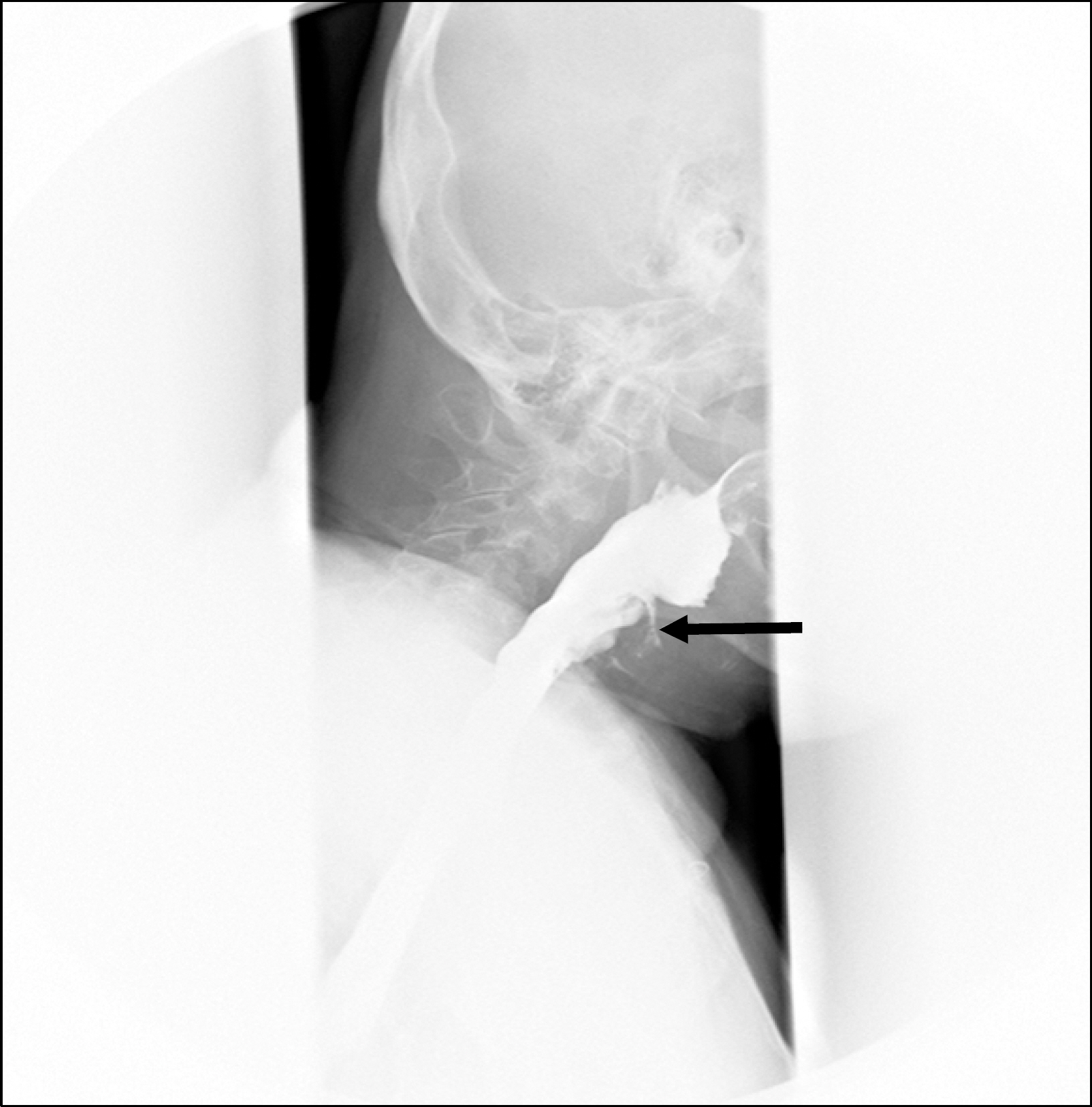
Fig. 2 Esophagogram was taken 13 days after operation. Arrow showed esophageal leak.
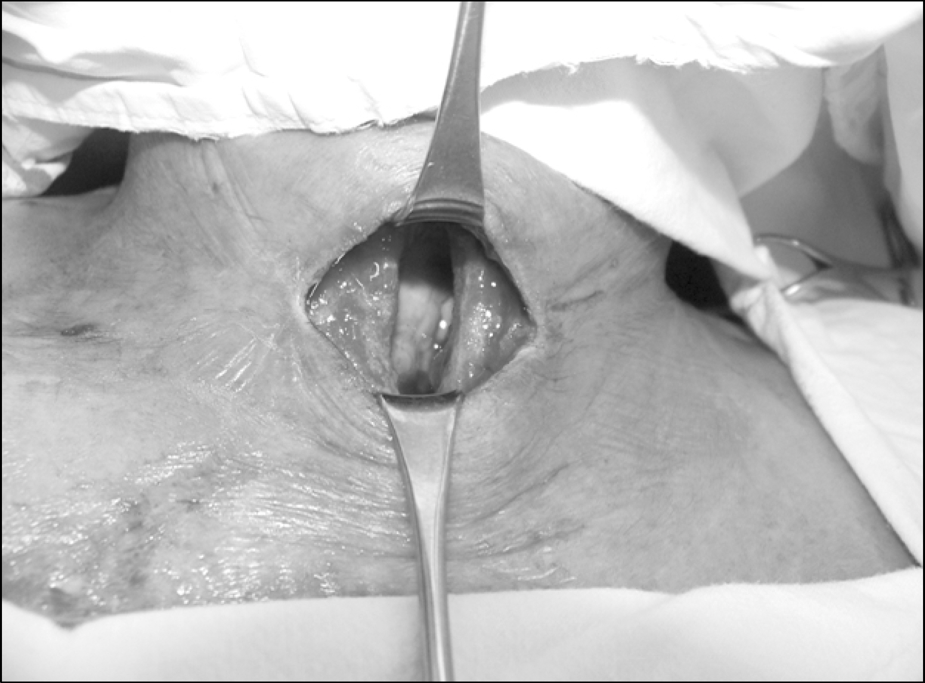
Fig. 3 Trachea and perforation site, still exposed, 14 days after operation.
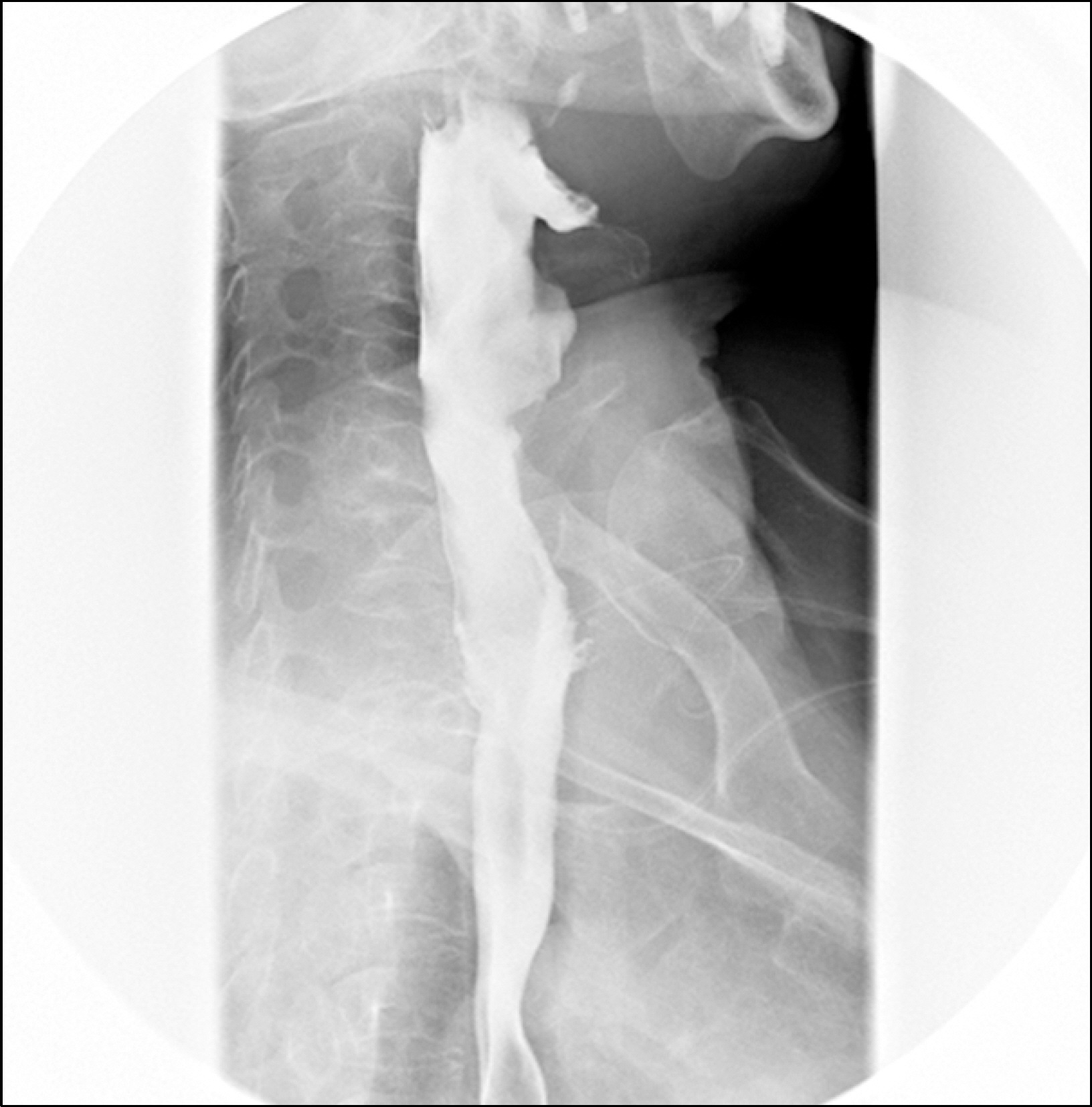
Fig. 4 Esophagogram on 18th postoperative day showed no evidence of leak.
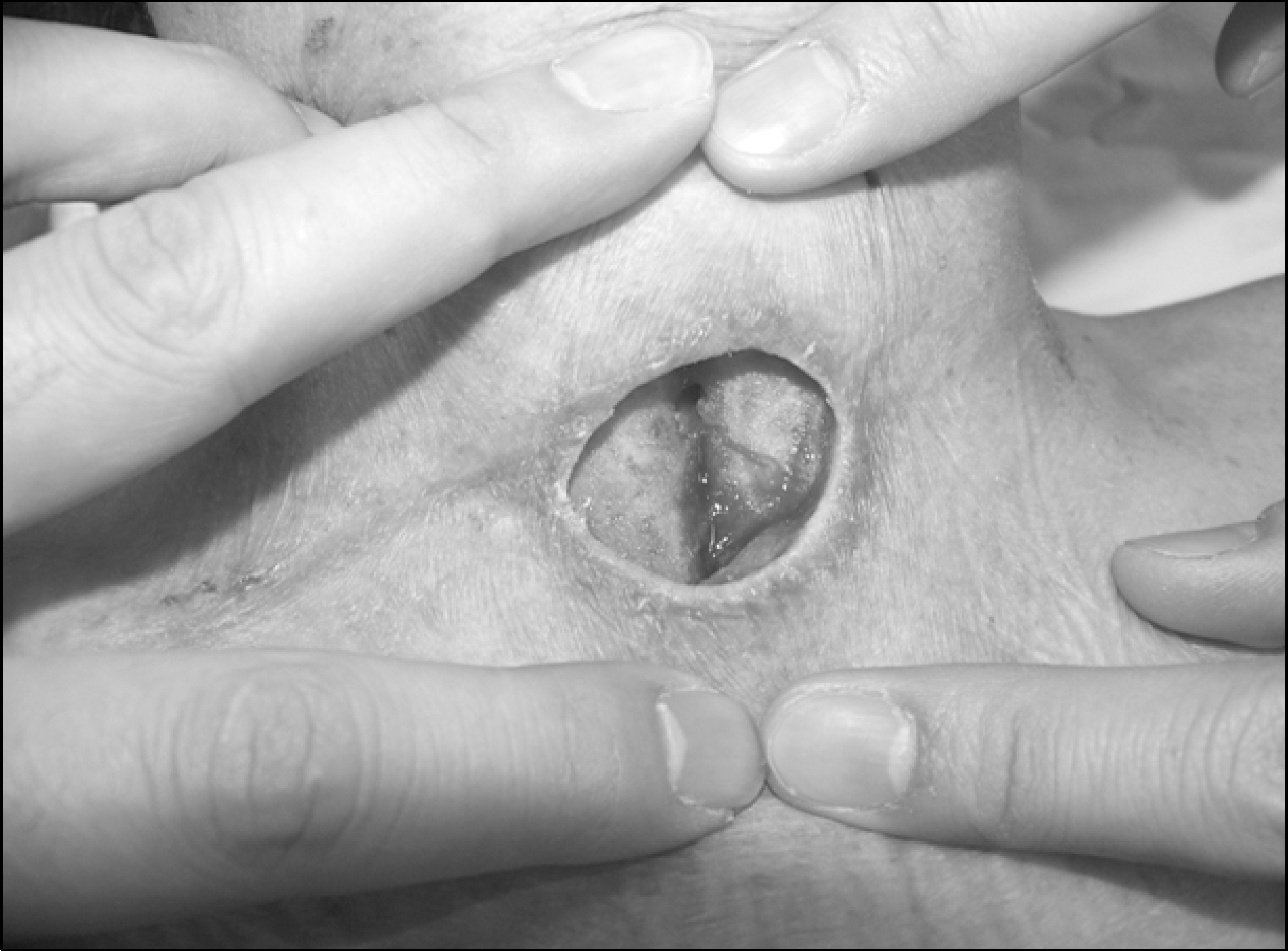
Fig. 5 Granulation tissue, 18 days after operation, covered the trachea and grew upto subcutaneous layer.
Reference
-
1.Bergamaschi R., Becouarn G., Ronceray J., Arnaud JP. Morbidity of thyroid surgery. Am J Surg. 1998. 176:71–5.
Article2.Nikolaos ND., Apostolakis EE., Marangos MN., Koletsis EN., Zampakis P., Panagopoulos K, et al. A less invasive management of post-thyroidectomy descending necrotizing mediastinitis is feasible: a case report and literature review. Med Sci Monit. 2007. 13:CS83–7.3.Madanick RD. Medical management of iatrogenic esophageal perforations. Current Treatment Options in Gastroenterology. 2008. 11:54–63.
Article4.Fleischmann W., Strecker W., Bombelli M., Kinzl L. Vacuum sealing as a treatment of soft tissue damage in open fractures. Unfallchirurg. 1993. 96:488–92.5.Fleischmann W., Lang E., Klinzl L. Vacuum assisted wound closure after dermatofasciotomy of the lower extremity. Unfallchirurg. 1996. 99:283–7.6.Dhir K., Reino AJ., Lipana J. Vacuum-assisted closure therapy in the management of head and neck wounds. Laryngoscope. 2009. 119:54–61.
Article7.Morykwas MJ., Argenta LC., El Shelton-Brown., McGuirt W. Vacuum-assisted closure: a new method for wound control and treatment: animal studies and basic foundation. Ann Plast Surg. 1997. 38:553–62.8.Saxena V., Hwang CW., Huang S., Eichbaum Q., Ingber D., Orgill DP. Vacuum-assisted closure: microdeformations of wounds and cell proliferation. Plast Reconstr Surg. 2004. 114:1086–96.
Article9.Argenta LC., Morykwas MJ. Vacuum-assisted closure: a new method for wound control and treatment: clinical experience. Ann Plast Surg. 1997. 38:563–77.10.Venturi MA., Attinger CE., Mesbahi AN., Hess CL., Graw KS. Mechanisms and clinical applications of the vacuumassisted closure (VAC) device: a review. Am J Clin Dermatol. 2005. 6:185–94.
- Full Text Links
-
- Actions
-
Cited
- CITED
-
- Close
- Share
-
- Similar articles
-
- Usefulness of Vacuum-Assisted Closure Therapy in the Treatment of Esophageal Perforation Following Thyroidectomy
- Endoscopic Vacuum-assisted Closure in a Patient with an Overtube-induced Esophageal Perforation
- Esophageal Endoscopic Vacuum Therapy with Enteral Feeding Using a Sengstaken-Blakemore Tube
- Successful Endoscopic Vacuum-Assisted Closure Therapy for Esophageal Perforation: A Case Report
- Esophageal perforation in children: etiology and management, with special reference to endoscopic esophageal perforation
